The fiber is thinner than a human hair, its tip coated with a film of gold so thin it barely counts as a layer at all. Aim a laser into it and something odd happens: the gold heats up, a tiny bubble blooms at the tip, and the liquid surrounding it begins to spin. Within 60 seconds, thousands of bacteria that were scattered invisibly through the sample have been swept together into a tight cluster, ready to be counted, identified, interrogated. The whole thing fits in your hand. This is optical condensation, and it may change how quickly dangerous bacteria get caught.
The challenge at the heart of disease detection has always been the same: harmful microbes are dilute, and dilute things are hard to find. E. coli O157, for instance, can trigger serious illness with as few as ten cells in a sample; conventional lab culture takes days to confirm its presence, and even faster immunoassay methods still demand several hours.
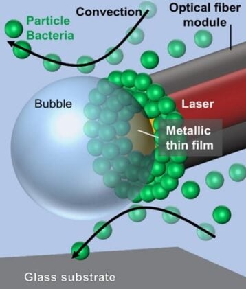
Researchers at Osaka Metropolitan University have now demonstrated a light-driven approach that sidesteps both problems. Their device, described in Communications Physics, uses a commercially available multimode optical fiber stripped of its polymer jacket and coated at the tip with a 10-nanometer gold film via ion sputtering. When an infrared laser is fed into the fiber, the gold film absorbs photons and converts them into heat through the photothermal effect. The local temperature spikes sharply enough to nucleate a microscopic bubble in the surrounding liquid. That bubble, it turns out, is the key to everything. Surface tension drops more steeply on the hot underside of the bubble than on its cooler upper surface, generating what physicists call Marangoni convection: a swirling, three-dimensional flow that draws fluid inward from all directions and funnels bacteria and particles into the stagnant zone between the bubble and the fiber tip, where they accumulate.
“Many conventional techniques are time consuming, require complex instrumentation, or are limited to collecting targets only near a surface or within a narrow focal region,” said Takuya Iida, professor at the Graduate School of Science and lead author of the study.
What makes this approach unusual is the geometry. Photothermal condensation is not itself new; earlier versions placed a gold-coated flat substrate at the base of a sample droplet and heated it with a focused laser from below. That worked, but it only pulled bacteria horizontally, along the surface, and substrate friction slowed the flow. The fiber hangs in the liquid at any chosen depth, and the resulting convection sweeps in from the sides and from above and below simultaneously. “Unlike conventional photothermal techniques that primarily operate in two dimensions along a surface, this system captures targets from all directions within the liquid,” Iida said.
The difference in efficiency is striking. Across a 20-microliter sample, the fiber module assembled somewhere between a thousand and a hundred thousand particles or bacteria within 60 seconds. Assembly efficiency (the fraction of all available objects actually collected) reached roughly 10 percent at low concentrations. The conventional flat-substrate approach topped out around 0.9 percent under the same conditions; the fiber version exceeded it by more than tenfold.
Bacteria Corralled, With Caveats
The team tested the system on live E. coli, stained with fluorescent dyes that distinguish living cells from dead ones. Bacteria assembled at the fiber tip just as the polystyrene test particles had, though in somewhat smaller numbers; their irregular rod shapes and surface chemistry make them harder to corral than smooth spheres. Assembly efficiency for bacteria ran between about 7 and 10 percent depending on concentration, which is comfortably above any previous substrate-based result. The complication: the laser heat killed most of them. At the concentrations tested, fewer than half the assembled bacteria survived the process. For a diagnostic application that only needs to detect and count cells, viability may not matter. But any use case requiring live bacteria, including tests of antibiotic susceptibility, will need a lower-temperature solution. The paper suggests plasmonic nanostructures or honeycomb geometries at the fiber tip could localise heat more precisely and reduce collateral damage; it is a known problem in this field, not a surprise.
The researchers also demonstrated collection of polystyrene nanoparticles 100 nanometers across, well below the diffraction limit where optical tweezers typically struggle. Assembly efficiency for those was lower (below 1 percent), but still significantly above previous benchmarks.
Small Sample, Big Ambition
“Our results demonstrated that complex optical setups are not required to achieve high-efficiency concentration, and that a compact fiber-based approach can substantially enhance collection performance in liquid environments,” Iida said. That compactness matters as much as the performance numbers. Conventional optical condensation rigs require objective lenses, precisely positioned substrates, and a fairly rigid optical bench. The fiber module, by contrast, can in principle be dipped into any small liquid volume and interrogated at any depth, which opens possibilities for field diagnostics, endoscopic applications, and environmental sampling where fixed lab geometry is not an option.
The immediate next step is integration. Optical condensation on its own concentrates bacteria; it doesn’t identify them. Combining the fiber module with surface-enhanced Raman spectroscopy or fluorescence-based immunoassays would let the concentrated cluster be interrogated in place, within the same 60-second window. The team has also flagged the possibility of coupling the approach with antigen-antibody reactions, which would allow selective capture of specific pathogens from complex mixtures.
“Ultimately, we aim to develop a versatile and reliable approach for rapid, sensitive analysis in small-volume liquid samples, contributing to future advances in bioanalytical research, environmental monitoring, and related analytical technologies,” Iida said. Whether that ambition translates into a deployable diagnostic tool depends largely on whether the bacterial survival problem can be cracked. But the geometry is sound, the efficiency gains are real, and the hardware is, by the standards of optics labs, refreshingly simple. Sometimes a fiber, a bit of gold, and the physics of a bubble is enough.
Frequently Asked Questions
Why is it so hard to detect dangerous bacteria quickly?
The core problem is concentration: harmful bacteria like E. coli O157 can cause serious illness at extremely low numbers, meaning they’re often too sparse in a sample to detect easily. Traditional culture methods grow colonies over days, while faster antibody-based tests still take several hours. Any technique that first gathers bacteria into a tight cluster before analysis dramatically shortens both the time and the detection threshold required.
How does a bubble help collect bacteria?
When the laser heats the gold tip, it nucleates a tiny bubble in the liquid. The bubble’s surface is hotter at the bottom (near the fiber) and cooler at the top, creating a surface tension gradient. That gradient drives Marangoni convection, a swirling fluid flow that sweeps inward from all directions and funnels particles into the calm zone between the bubble and the fiber tip, where they accumulate. It’s less like a magnet and more like a microscale whirlpool with a quiet eye.
Could this work outside a laboratory?
That’s the aim. Because the fiber can be dipped into any small liquid sample at any depth, the setup doesn’t require the rigid, precisely aligned optics of conventional lab instruments. The researchers flag potential applications in field diagnostics, environmental monitoring, and even endoscopy, where collecting and concentrating biological targets in situ would be valuable. A fully portable version would require further miniaturisation, but the hardware is already far simpler than competing approaches.
What stops it from being used for diagnosis right now?
Two things. First, the laser heat kills a significant proportion of collected bacteria, which limits any application requiring live cells. Second, optical condensation on its own only concentrates organisms; it doesn’t identify which species or strain is present. The team plans to integrate the fiber module with downstream analytical tools like Raman spectroscopy or immunoassays. Once concentration and identification can be done in the same 60-second window, something close to a deployable diagnostic tool becomes plausible.
https://doi.org/10.1038/s42005-025-02480-9
ScienceBlog.com has no paywalls, no sponsored content, and no agenda beyond getting the science right. Every story here is written to inform, not to impress an advertiser or push a point of view.
Good science journalism takes time — reading the papers, checking the claims, finding researchers who can put findings in context. We do that work because we think it matters.
If you find this site useful, consider supporting it with a donation. Even a few dollars a month helps keep the coverage independent and free for everyone.