Hidden inside an opaque plastic cup, suspended in clear gelatin, sits a small metal object: maybe a spring, maybe a screw, maybe a ball bearing. Your job is to figure out which, using nothing but sound. Slide a probe across the surface, watch the grainy slices flicker on a monitor, and try to assemble a shape in your head from a stack of two-dimensional shadows. For a trained sonographer, this is routine. For a beginner, it is genuinely hard, and that gap between the two is exactly what a team at MIT set out to close.
Their answer was to stop asking people to do the assembling at all. Instead of slices on a screen, they built a system that streams a live three-dimensional rendering of whatever the probe is looking at straight into an augmented-reality headset, parked in mid-air over the real object like a ghostly cutaway.
The trouble with conventional ultrasound has always been the translation step. Sound waves bounce off tissue, return to the transducer, and get converted into a flat image: one plane, one angle, one slice of a body that is emphatically not flat. The operator has to mentally stack those slices into something solid. Jason Hou, an MIT graduate student and lead author of the work, calls it the central obstacle of the whole discipline. “The hardest thing is this mental tomography bottleneck where you’re trained to reconstruct the 2D slices in your 3D mental space. That is a cognitive burden that can lead to inaccuracies in scanning,” he says.
And it is not a small burden. “It’s a difficult skill to master, and there are long learning curves,” Hou says. People spend years getting good at it.
X-ray vision, more or less
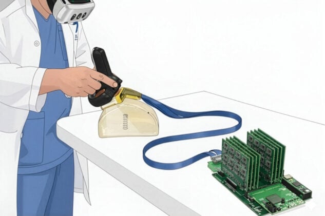
The MIT system, which the team calls AR-VIU (for augmented real-time volumetric imaging in ultrasound), pairs two technologies that don’t usually travel together. One is a compact 3D ultrasound probe, slightly smaller than a deck of cards, built around an unusual square-shaped array that lets it capture volumes directly rather than reconstructing them after the fact. The other is a mixed-reality headset. Voxel data from the probe gets compressed, streamed into Unreal Engine (the same graphics software that powers video games), and rebuilt as a point cloud you can walk around.
The effect, when you put the headset on, is something close to X-ray vision. The rendering hovers over the physical object’s actual location, and by tilting your head or stepping to one side you see it from a new angle, the way you would inspect any real thing sitting on a table. No mental gymnastics required. The structure is just, well, there.
To find out whether that actually helped, the researchers ran 18 people through a set of identification and localization tasks. Nine were experts, sonographers and physicians who scan for a living. Nine had never touched an ultrasound probe before. Everyone tried four setups: conventional 2D on a screen, 3D on a screen, 2D in augmented reality, and the full 3D-in-AR combination. In one task they identified those hidden gelatin-trapped objects. In another they marked, with a pen, where a target sat inside a block of tissue-mimicking gel, standing in for the delicate business of guiding a biopsy needle to the right spot. The numbers came out lopsided in AR-VIU’s favor. Object identification accuracy hit about 92 per cent, and the system gave users roughly eight times the odds of getting it right compared with the standard flat-screen setup.
The more striking result was about who improved. Under conventional 2D imaging, experts ran rings around the novices, as you’d expect. Switch to AR-VIU and that gap essentially vanished: beginners performed nearly as well as the veterans. Novice targeting error dropped by more than half. “Overlaying images with the anatomy and providing 3D visual context makes ultrasound significantly easier for novices to understand,” says Shrihari Viswanath, another lead author.
The experts weren’t sold
Here’s the wrinkle, though. The experts, by and large, didn’t want it. Most of them preferred the old 2D screen, and not out of stubbornness so much as hard-won competence: they had spent years building exactly the mental machinery the new system makes unnecessary, and they were quick on the flat plane. As one expert, a cardiologist with more than a decade of echocardiography behind them, put it in a post-study interview: “I preferred [2D] just because that’s what I was used to, and I could just refer to it on a flat plane.” A skill that took years to acquire is not lightly set aside.
Even so, the same experts could see where the thing would earn its keep. About two-thirds of them volunteered specific clinical uses without being prompted: vascular access, biopsy guidance, trauma assessment, watching a heart wall move during echocardiography. One sonographer, noting that in conventional scanning “the operator dependence is very heavy,” pointed to exactly the situations where a less experienced hand needs all the spatial context it can get.
For now the system is far from a hospital fixture. It runs at about two volumes per second, the headset is heavy, and the resolution still trails what a good conventional machine delivers, all of which several participants flagged. Senior author Canan Dagdeviren frames the near-term promise mostly in terms of teaching and confidence, where the system “could make ultrasound more intuitive and more understandable,” and on the clinical side “less time-consuming, more accurate,” sparing providers the nagging worry that they missed something. The researchers are now pushing on resolution and accuracy. Whether AR-VIU ever ends up in an emergency room is an open question, but it has already shown something quieter and more interesting: that a stubborn human bottleneck, the one that takes years of practice to clear, might instead be handed off to a machine that simply shows you the shape.
Hou, J.F., Viswanath, S., Dilibal, C. et al. Communications Engineering 5, 107 (2026).
Frequently Asked Questions
Why is reading a normal ultrasound so hard to learn?
A standard ultrasound shows flat, two-dimensional slices, but the body it’s imaging is three-dimensional. The operator has to mentally stack those slices into a solid shape in real time, a skill researchers call the “mental tomography bottleneck” that can take years of practice to master. Reducing that cognitive load is the whole point of the new approach.
How does projecting ultrasound into a headset actually help?
Instead of asking the user to reconstruct a 3D shape from 2D images in their head, the system renders the scanned object as a live 3D image floating over its real location, viewable from any angle through an AR headset. That removes the translation step entirely, letting beginners see structure directly rather than infer it. The result was a near-elimination of the gap between novice and expert performance.
Could this replace conventional ultrasound machines?
Not yet, and maybe not entirely. The current system runs slowly, the headset is bulky, and image resolution still lags behind standard equipment, so researchers see its nearest-term value in training and in specific procedures like biopsy guidance. Experienced sonographers in the study still preferred the familiar flat screen they had trained on for years.
Why did expert users prefer the old 2D screens?
Experts have already built the exact mental skill the new system makes unnecessary, so for them the flat screen is fast and trusted rather than burdensome. Several still acknowledged that the 3D headset would help in cases demanding quick spatial judgment, such as placing a needle or watching the heart wall move. The benefit, in other words, depends heavily on who is holding the probe.
ScienceBlog.com has no paywalls, no sponsored content, and no agenda beyond getting the science right. Every story here is written to inform, not to impress an advertiser or push a point of view.
Good science journalism takes time — reading the papers, checking the claims, finding researchers who can put findings in context. We do that work because we think it matters.
If you find this site useful, consider supporting it with a donation. Even a few dollars a month helps keep the coverage independent and free for everyone.