In Madrid, a scientist named Santiago Ramón y Cajal looks through a microscope at a blackened piece of brain and, for the first time, saw single nerve cells with every branch visible. He was “enraptured,” as the Nobel Prize’s history of Cajal puts it.
The year was 1887, but to get the full story, we have to go back to 1872, when a young Italian doctor, short on money, took a job at a hospital outside Milan. He built a lab in a kitchen, and the research he did there would go on to win him a Nobel Prize some 33 years later.
A hospital for incurables and a lab in the kitchen
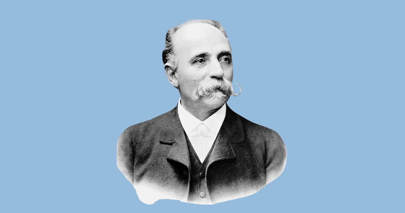
The doctor was Camillo Golgi. He had a “solid reputation” in Pavia, but no secure university job and not much money.
Pressured by his father, he became chief physician at a hospital for chronic illness in the small town of Abbiategrasso. His biographer, Paolo Mazzarello, writes that “everything indicated” his “research activities were about to end for good.”
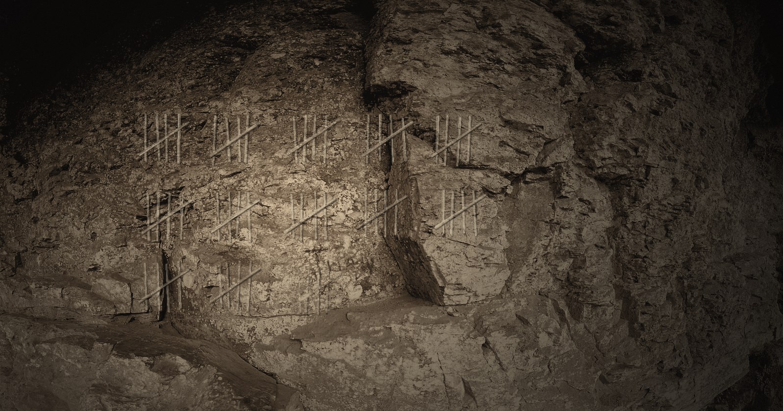
They didn’t. He turned the kitchen of his quarters into a small lab, with a microscope and a few instruments. The hospital’s twice-monthly post-mortems gave him tissue to study, and he worked at the microscope by candlelight.
In those days, brain tissue under a microscope was close to unreadable. The usual stains gave a poor picture. You could see that cells were there, but not where one ended and the next began. Golgi was searching for a better stain, and his answer combined two chemicals he had already used.
First, you harden a block of brain in potassium dichromate for days or weeks. Then you soak it in silver nitrate. Crystals form inside a few scattered cells and turn them black, right out to their finest branches. Because the rest stay pale, each blackened cell stands out on its own.
On February 16, 1873, he wrote to a friend:
“I am delighted that I have found a new reaction to demonstrate, even to the blind, the structure of the interstitial stroma of the cerebral cortex.”
The “interstitial stroma” is the brain’s supporting tissue, so he wasn’t yet claiming to see whole nerve cells. He published the method that year in a short note in a Lombard medical journal.
A discovery slow to spread
For more than a decade, few others took notice but Golgi kept at it. He returned to Pavia as a professor in 1876 and spent about ten years staining different parts of the brain, work he collected in a book in 1885.
He saw things nobody had seen before. A nerve cell’s long output fiber, the axon, sprouted fine side branches. Its shorter branches, the dendrites, ended freely instead of joining up into a web.
Still, he didn’t let go of the leading theory of the day, which held that the nervous system was one continuous network. Golgi built his own: a “diffuse nervous net” in which the axons were connected.
So why did the method not take off in the scientific community?
First of all, as noted by the Science History Institute, it appeared in a journal with limited circulation, with no pictures.
But the bigger problem, as the neuroscientist Edward G. Jones explains, was that few scientists could make it work. Even the eminent Swiss histologist Albert von Kölliker visited Pavia in 1888 and failed.
By then Golgi had moved on to studying malaria.
Madrid, 1887: “a look was enough”
This brings us back to Madrid.
At the time, Cajal was 35 and a professor of anatomy in Valencia. A psychiatrist named Luis Simarro, just back from Paris, showed him some Golgi-stained slides.
In his autobiography, Cajal described cells “coloured brownish black even to their finest branchlets, standing out with unsurpassable clarity upon a transparent yellow background. All was sharp as a sketch with Chinese ink.”
By one account, he couldn’t sleep that night and was back at Simarro’s door the next morning. Within a year he had dropped all his other work.
Cajal then found ways to make the method more reliable. He repeated the staining, soaked tissue longer in stronger solutions, and used young animals whose nerve fibers weren’t yet heavily coated in myelin. After that, others could make it work too.
Cajal published in a journal he paid for himself, and it used up all his savings.
What he saw changed the argument. In the cerebellum and the retina, nerve endings reached toward other cells and stopped. So nerve cells, he argued, were separate units that communicate by contact. He never saw a gap directly. He read it into the blank spaces where the stain stopped.
By 1891 the idea had a name: the neuron doctrine.
The two things Golgi got right, branching axons and free-ending dendrites, became part of Cajal’s case against him. In 1889 Cajal took his slides to Berlin, and Kölliker became a supporter.
Cajal also wrote to Golgi that his preparations were so clear that doubts about them were “absurd.” Golgi sent back a paper on malaria.
Stockholm, 1906: one prize, two nervous systems
The two men didn’t meet until December 1906, in Stockholm, when they shared the Nobel Prize “in recognition of their work on the structure of the nervous system.”
The vote for a shared prize wasn’t unanimous. The Swedish anatomist Gustaf Retzius, a member of the Nobel committee, summed up the pair: Golgi supplied “the key to open the building that encloses the secrets of the nervous system, but only Ramón y Cajal taught us how to use it.”
Golgi lectured first, on December 11. He was a modest, reserved man, which makes his opening line all the more surprising: “It may seem strange that, since I have always been opposed to the neuron theory … I have chosen this question of the neuron as the subject of my lecture, and that it comes at a time when this doctrine is generally recognized to be going out of favour.”
It wasn’t going out of favor. A sentence later, he admitted that “the majority of physiologists, anatomists and pathologists still support the neuron theory.”
He defended his diffuse net and closed: “I cannot abandon the idea of a unitary action of the nervous system, without bothering if, by that, I approach old conceptions.”
Cajal spoke the next day. He was polite, crediting “my celebrated colleague Professor Golgi,” but he made his point:
“Unfortunately, nature seems unaware of our intellectual need for convenience and unity, and very often takes delight in complication and diversity.”
In private, he was furious. He later recalled sitting through Golgi’s lecture “trembling with impatience,” and called it “a cruel irony of fate” to pair, “like Siamese twins united by the shoulders, scientific adversaries of such contrasting character!”
The lecture badly damaged Golgi’s reputation.
Why does it stain only a few cells?
Golgi may have lost the argument, but his stain is still in use. Its strength is that it stains only a few cells, usually 1% to 5%, and leaves the rest alone.
Why those cells and not the others? Nobody knows.
Its weakness is reliability. As noted by Rafael Yuste, the method is still “notoriously capricious,” and how it works is poorly understood.
Even so, it is still in use more than 150 years on, with a 2014 study calling it “the predominant method for viewing the dendritic structure of human neurons”.
Pavia, 2019: still black under the microscope
Most of Golgi’s slides were lost after his death but 38 survive in two museums in Pavia, and six have labels signed by Golgi.
In a 2019 study, researchers looked at the slides under a modern microscope. Two sections they measured were 150 to 200 microns thick, so thick they needed constant refocusing.
And the spines were there, “especially evident” on some cortical neurons. The team’s best guess is that Golgi left them out because “he believed that they could be artifacts.”
He finally drew them in 1901, without comment. The structures he hadn’t recognized were there all along in preparations from his own laboratory.
In 1887 Cajal saw black cells on “a transparent yellow background.” More than 130 years later, the 2019 team described Golgi’s own slides in almost the same words: “The impregnated cells stand out in black over a light yellow background.”
Looking at them, they wrote, “it is unavoidable to guess the surprise of being able to observe nerve cells in their entirety for the first time.”
That surprise started in a kitchen in Abbiategrasso.