Scientists have developed a flexible, conductive hydrogel that may significantly advance how researchers monitor spinal cord activity during movement, potentially transforming future approaches to spinal injury research and treatment.
The research team, led by Assistant Professor Siyuan Rao from Binghamton University’s Thomas J. Watson College of Engineering and Applied Science, created a novel electrode design that addresses a critical challenge in neural interfaces: maintaining electrical connection while accommodating the natural movements of tissues.
The new approach creates a motion-adaptive soft-material neural interface that minimizes tissue damage during animal movement while still capturing high-quality physiological signals. As Rao explained in the press release, “If you have a rigid material in a soft tissue, especially during movement, it’s going to cause a lot of damage. Our technology solves that fundamental problem, so we can pick up a single cell’s activity from the spinal cord and maintain the device’s functionality for a long time.”
The technology, which the researchers call TRAIN (Tension Reinforcement for Anisotropic Nano-orientation), uses carbon nanotubes embedded within polyvinyl alcohol hydrogels. These microscopic carbon structures form electrically conductive pathways within the gel, allowing the transmission of neural signals while maintaining the material’s flexibility.
What distinguishes this approach is how the team manipulates the material’s internal structure. By repeatedly stretching the hydrogel in a specific direction, they cause the embedded carbon nanotubes to self-align, creating directional conductivity—much like training muscle fibers to strengthen in a particular orientation.
The cyclic stretching process systematically realigns the nanotubes along the stretching direction. This creates preferred electrical pathways that improve conductivity precisely in the direction needed, while the material remains soft and tissue-compatible.
In laboratory tests, the hydrogel electrodes demonstrated remarkable durability, withstanding up to 20,000 stretching cycles at 20% strain—roughly simulating the repeated movements tissues might experience in daily activity. When implanted in mice, the electrodes successfully recorded both muscle activity and spinal neuron signals over extended periods.
One notable achievement was the electrode’s ability to function for eight months after implantation in the ventral spinal cord of mice—a significant improvement over conventional technologies that often fail due to mechanical stress or tissue rejection.
The researchers were able to record single neuron activity from deep within the spinal cord over this extended timeframe. This is particularly valuable because accessing these deeper ventral structures, which control motor function, has been extremely challenging with traditional surface electrodes.
The hydrogel’s composition resembles a specialized sponge that contains water and nearly invisible carbon nanotubes distributed throughout its three-dimensional network. As Huang described it, “You can imagine it as a sponge that contains a lot of water and the conductive material, which are the nanocarbon tubes invisible to the naked eye because they’re so small.”
This structure closely mimics the mechanical properties of actual neural tissue, with an elasticity of approximately 16 MPa—far softer than conventional metal or silicon electrodes.
Importantly, mice implanted with the electrodes showed no significant differences in their movement patterns compared to control animals, suggesting the technology integrates well with natural biological functions.
The research builds on growing interest in “soft electronics” for biomedical applications, where traditional rigid materials often trigger inflammation, scarring, and signal degradation over time—problems that have limited the long-term viability of neural interfaces.
The team used advanced imaging techniques to verify the alignment of carbon nanotubes within the hydrogel. Using stimulated Raman scattering microscopy, they could directly visualize how the carbon nanotubes reorganized after the stretching process, confirming their hypothesis about the mechanism behind the improved directional conductivity.
The implications extend beyond basic research tools. Similar hydrogel technologies could eventually be adapted for therapeutic applications in humans with spinal cord injuries or neurological disorders, though such clinical applications would require years of additional development and safety testing.
Rao’s team is now focusing on expanding the technology’s capabilities. They are particularly interested in combining this with optogenetic approaches that could potentially inhibit pain signals or stimulate motor function recovery. As Rao noted, “We specifically want to look at the ventral horn motor neurons that control voluntary movement.”
The study reflects a growing trend toward interdisciplinary collaboration in neurotechnology, with contributors from multiple institutions including the University of Massachusetts, the University of Texas, Michigan State University, MIT, and Boston Children’s Hospital.
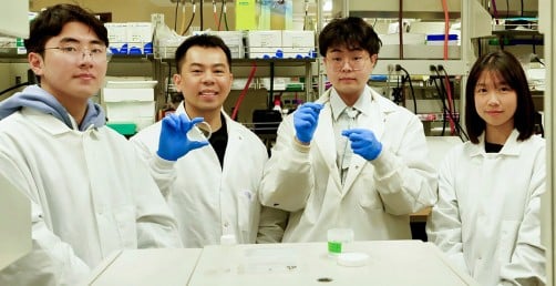
For Huang, who supervised several students during the project, the research provided valuable leadership experience. “It’s like we’re in a car,” he said. “I’m in the driver’s seat, and all of the master’s students are my passengers — but they don’t just sit in the car. They also contribute, like one of them is keeping an eye on the maps to tell me directions, or I tell them directions. That’s how we work.”
As neural interface technologies continue to advance, this hydrogel approach represents a promising direction for creating durable connections between electronic devices and living tissue—addressing one of the most persistent challenges in bioelectronics.
The spinal cord experiences complex mechanical dynamics during natural movement. This new technology adapts to these conditions rather than fighting against them, which explains how the researchers have been able to maintain stable recordings over such extended periods.
If our reporting has informed or inspired you, please consider making a donation. Every contribution, no matter the size, empowers us to continue delivering accurate, engaging, and trustworthy science and medical news. Independent journalism requires time, effort, and resources—your support ensures we can keep uncovering the stories that matter most to you.
Join us in making knowledge accessible and impactful. Thank you for standing with us!