Researchers at UC Irvine have developed an imaging method that captures detailed 3D images using just one X-ray exposure, potentially reducing radiation exposure and eliminating the need for bulky CT scanning equipment. The technique, called X-ray-Induced Acoustic Computed Tomography (XACT), could make advanced medical imaging more accessible worldwide.
Published in Science Advances | Estimated reading time: 6 minutes
Sound Waves Unlock a New Dimension
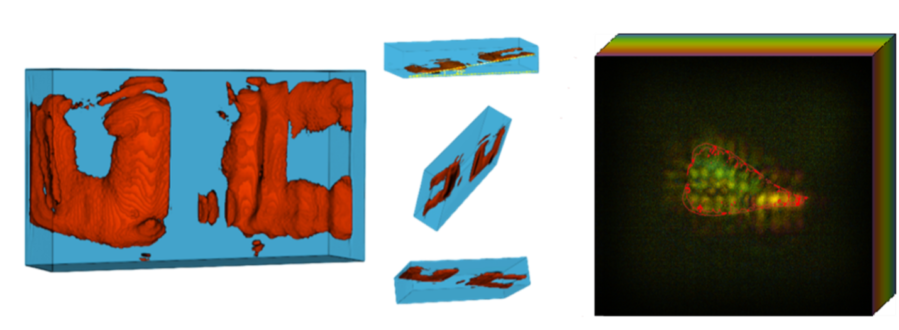
The research team, led by Associate Professor Shawn Xiang, found a way to convert X-ray energy into sound waves that provide three-dimensional information. “In XACT, the generated sound waves by X-rays change the way X-ray imaging works, converting X-rays to ultrasound,” explains Xiang. “X-rays typically travel in straight lines, so one projection only provides 2D information. However, X-ray-induced acoustic signals propagate in three dimensions, allowing for 3D imaging with a single projection.”
Breaking Through Traditional Limitations
Traditional CT scanners require hundreds of X-ray images taken from different angles, exposing patients to significant radiation and requiring large, immobile equipment. XACT captures acoustic waves traveling at 1,500 meters per second through tissue, allowing real-time 3D visualization with a single exposure. The team achieved resolution of 0.4 millimeters in one plane and 3.5 millimeters in another, demonstrating the technology’s potential for detailed medical imaging.
From Laboratory to Clinical Practice
The implications for healthcare access are significant. XACT’s compact design and reduced radiation exposure could make advanced imaging available in settings previously unable to support traditional CT scanners, including rural areas, battlefield locations, and even space stations. The research team has already demonstrated the technology’s effectiveness on both synthetic materials and biological tissue.
Glossary
- XACT (X-ray-Induced Acoustic Computed Tomography): A technique that converts X-ray energy into sound waves for 3D imaging
- Acoustic Wave: A sound wave generated by X-ray interaction with tissue that travels at 1,500 meters per second
- Resolution: The ability to distinguish between small, closely spaced objects in an image
Test Your Knowledge
What is the main advantage of XACT over traditional CT scanning?
XACT can create 3D images with a single X-ray projection, while traditional CT requires hundreds of projections from different angles.
How fast do the acoustic waves travel in XACT imaging?
The acoustic waves travel at 1,500 meters per second.
What resolution did researchers achieve with XACT?
The system achieved resolutions of 0.4 millimeters in the XZ plane and 3.5 millimeters in the XY plane.
How does XACT’s mechanism of converting X-rays to acoustic waves enable 3D imaging?
While X-rays travel in straight lines providing only 2D information, the X-ray-induced acoustic signals propagate in three dimensions, allowing for 3D imaging with a single projection.
Enjoy this story? Subscribe to our newsletter at scienceblog.substack.com.
ScienceBlog.com has no paywalls, no sponsored content, and no agenda beyond getting the science right. Every story here is written to inform, not to impress an advertiser or push a point of view.
Good science journalism takes time — reading the papers, checking the claims, finding researchers who can put findings in context. We do that work because we think it matters.
If you find this site useful, consider supporting it with a donation. Even a few dollars a month helps keep the coverage independent and free for everyone.