Researchers at the Mark and Mary Stevens Neuroimaging and Informatics Institute at the Keck School of Medicine of USC have found a new pattern in one of the brain’s key memory hubs. In mice, they discovered that the CA1 region of the hippocampus is built from four distinct layers of nerve cells, each defined by its own gene activity.
The study in Nature Communications could help explain how different parts of CA1 support different behaviors and why some cells are hit harder in conditions like Alzheimer’s disease and epilepsy.
The hippocampus is a deep brain structure or “memory gateway” that is crucial for forming new memories and mapping space. CA1 sits near the end of this circuit and acts as a major output station, passing processed information to regions involved in thinking and emotion. Understanding how its cells are arranged helps link brain wiring to behavior.
Four Layers Along A Memory Highway
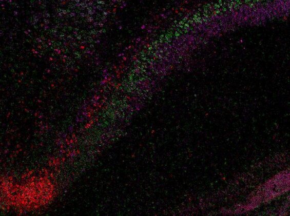
To reveal CA1’s hidden structure, the team used an RNA labeling technique called RNAscope with high resolution microscopy. This let them see single molecules of RNA in thin slices of mouse brain and identify which genes were active in each cell. Across 58,065 CA1 pyramidal neurons, they mapped more than 330,000 RNA molecules and grouped cells into types based on those patterns.
They found four continuous layers of pyramidal cells, stacked like thin sheets that run along the length of the hippocampus. Each layer has a distinct gene signature and changes slightly in thickness and position from one end of CA1 to the other. This layered view helps make sense of earlier work on sublayers and brain rhythms, including research on how cells switch between different brain rhythms to support learning and memory.
Clarifying A Tricky Border
The study also clarifies the border between CA1 and the subiculum, a neighboring region tied to memory and navigation. Under a microscope, this junction can look like a jumble of overlapping layers, making it hard to tell where CA1 ends and the subiculum begins. By labeling marker genes for both regions in the same tissue sections, the team could see how they meet.
They observed a gradual transition near the tail end of the hippocampus: the deepest CA1 layer fades while subiculum layers become more prominent. In this zone, cells from both regions sit side by side, which explains the complex, stacked appearance seen in atlases. A clearer border will help researchers interpret studies that link this junction to memory processing and seizure pathways, including work on brain hyperexcitability in epilepsy.
Why The Layers Matter For Disease
CA1 is one of the first hippocampal areas to degenerate in Alzheimer’s disease. This helps explain why patients forget loved ones and lose recent memories. CA1 is also part of the networks involved in temporal lobe epilepsy and mood disorders. By defining four precise CA1 layers with distinct gene signatures, the work offers a roadmap to test which neuron types are most vulnerable and how damage spreads.
If a disease process targets one layer, its impact will vary depending on where that layer is thickest and which regions it connects to. That may explain why some patients have more trouble with navigation while others struggle with recalling events or regulating emotions. It also fits with broader research on how the brain stores and backs up memories, suggesting layered circuits may add redundancy and flexibility.
Open Atlas For Future Studies
The CA1 cell type atlas builds on the Hippocampus Gene Expression Atlas and is freely available to researchers. It includes interactive 3D views accessible through the Schol-AR augmented reality app developed at the Stevens Institute, letting users explore how the layers run along CA1. These tools complement public resources on brain mapping and memory that connect lab data to big-picture neuroscience.
Because the pattern in mice resembles changes seen in primate and human CA1, similar laminar organization may be common across mammals. Confirming that will require studies in human and nonhuman primate tissue, but the new map already offers a clear framework. It links specific neuron layers to memory, navigation, and emotion, and points to concrete targets for therapies aimed at protecting vulnerable cells in Alzheimer’s disease and epilepsy.
“Our study shows that CA1 neurons are organized into four thin, continuous bands, each representing a different neuron type defined by a unique molecular signature,” Michael S. Bienkowski said.
“It’s like lifting a veil on the brain’s internal architecture. These hidden layers may explain differences in how hippocampal circuits support learning and memory,” Maricarmen Pachicano said.
ScienceBlog.com has no paywalls, no sponsored content, and no agenda beyond getting the science right. Every story here is written to inform, not to impress an advertiser or push a point of view.
Good science journalism takes time — reading the papers, checking the claims, finding researchers who can put findings in context. We do that work because we think it matters.
If you find this site useful, consider supporting it with a donation. Even a few dollars a month helps keep the coverage independent and free for everyone.