Nogo-A has been getting in the way for thirty years. The protein sits in the sheaths of nerve fibers throughout the spinal cord and brain, doing nothing obviously useful, yet doing something extremely effective: stopping damaged nerves from healing. After injury, when the body desperately needs axons to regrow and reconnect, Nogo-A is still there, blocking the process like a bouncer who doesn’t know the party’s over. Researchers at the University of Zurich have now shown, for the first time using live imaging, exactly how an antibody called NG101 removes that block, and what happens to the spinal cord when it does.
The findings matter because we have never, until now, been able to watch the effect of a regenerative treatment in a human spinal cord and actually see the tissue responding. That changes quite a lot of things.
Spinal cord injuries are profoundly cruel in a particular way. The damage itself may be brief, a collision, a fall, a bad dive, but the consequences cascade outward through months and years. The cord continues to shrink. Axons that were intact at the time of injury degenerate. Myelin erodes. The nervous system, which cannot regenerate itself the way skin or bone can, retreats rather than rebuilds. What the Zurich group has been pursuing, painstakingly, is a way to interrupt that retreat. NG101 was discovered at the university roughly three decades ago. It targets Nogo-A specifically, neutralizing it so that nerve fibers are no longer blocked from sprouting new connections. Clinical trials in patients with acute cervical injury began in 2019 under the multinational NISCI programme. Results published late in 2024 showed that the antibody was safe and appeared to improve upper extremity motor function. What nobody had established was the mechanism: not in humans, not in living tissue, not in real time.
Watching the Spinal Cord Rebuild
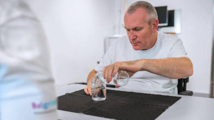
That’s what the new Nature Communications study provides. Using high-resolution MRI across 106 participants over six months, Patrick Freund’s team at Balgrist University Hospital tracked two parallel processes simultaneously. First, the lesion itself. Second, the cord above and below it.
The lesion findings were striking enough on their own. In patients receiving NG101, the volume of damaged tissue shrank by about 52 cubic millimetres per month. In the placebo group, it shrank by barely 2. More telling still was where the reduction was happening: primarily within the corticospinal tracts, the descending pathways that carry motor signals from the brain to the muscles. The dorsal columns, which carry sensory signals upward, showed similar rates of change in both groups. That tract-specific pattern rules out the obvious alternative explanation, that the treated patients were simply clearing edema faster. Widespread fluid resolution would affect everything equally. This didn’t.
The remote findings were perhaps more surprising. Even centimetres above the injury site, at the C1-C2 level of the cervical cord, the spinal cord in untreated patients was visibly atrophying, losing roughly 0.6 square millimetres of cross-sectional area every month. Standard Wallerian degeneration, the slow die-off of axons disconnected from their targets, proceeding exactly as expected. In the NG101 group that same measure was essentially flat. Myelin content, tracked using a technique called magnetization transfer saturation, showed an equivalent pattern: steady loss in controls, relative stability in treated participants. “We are now able to visualize the effect of the therapy early on and in an objective way,” Freund said. “This opens up the possibility of using future treatments more strategically and conducting a more reliable evaluation of their outcomes.”
What the imaging is probably capturing, the researchers believe, is a combination of things. Some regenerative sprouting around the lesion, new axons finding paths around the damaged area. Some protection of intact fibers that would otherwise have degenerated. The antibody, in Freund’s account, is doing both: slowing the retreat and encouraging a modest advance. “This allows surviving and newly regenerated nerve fibers to re-establish connections with the spinal cord centers that control the hand, arm and leg nerves,” he said. “These connections are essential for relaying signals from the brain to the muscles.”
The Signal Hidden in the Noise
One finding cuts against the grain in an instructive way. Among patients classified as motor-complete (meaning the injury had severed all voluntary motor function below the level of damage), NG101 produced structural changes similar to those seen in less severe cases: slower cord atrophy, better myelin preservation. But these patients showed no corresponding improvement in arm or hand function. None whatsoever. This might seem like a failure, but the researchers read it differently. The structural protection is happening even when the functional benefit isn’t measurable yet. Which suggests that behavioural outcomes, the scores and tests we use to assess recovery, are a fairly blunt instrument for detecting what’s actually going on in the cord.
That insight feeds directly into one of the study’s other main contributions: a new approach to clinical trial design. A persistent problem in spinal cord research is that patients vary enormously. A trial that mixes people with minor incomplete injuries alongside those with complete transection will have enormous variance in its outcomes, requiring very large samples to detect any treatment effect. The NISCI trial enrolled 126 patients over 39 months to reach statistical significance in its main outcome. The Zurich team showed that by combining two relatively simple screening measures, the width of preserved tissue at the injury site and the presence of detectable nerve signals in the legs, you can identify a subgroup of patients with much stronger and more consistent responses to NG101. In that subgroup, the required sample size to detect a significant functional improvement drops from 82 to 32. The enrollment period shortens by about a third. It’s not a small thing. Clinical trials in this population are already expensive, slow, and logistically complex. Cutting a third of the required recruitment time is, practically speaking, a different kind of result altogether.
There are caveats worth noting. The trial was mostly male, the MRI sub-cohort had baseline differences in lesion size between groups, and the stratification analysis was conducted retrospectively. These are genuine limitations, and the researchers acknowledge them directly. What they’re proposing is a framework for future trials, not a settled clinical algorithm.
Still. Something has shifted. We can now image a human spinal cord after acute injury, give a targeted biological treatment, and watch the tissue respond. Not infer it. Not model it. See it. “In our new study, we were able to use advanced imaging methods to show for the first time how this antibody therapy works directly in the spinal cord,” Freund said. Thirty years after Nogo-A was first identified in Zurich, the protein that blocks nerve repair has met something that stops it. The question now is how many people that can help, and whether imaging can tell us who, before the treatment even begins.
https://doi.org/10.1038/s41467-026-71412-0
Frequently Asked Questions
Can the NG101 antibody actually restore movement to paralysed patients?
The trial results suggest meaningful improvements in arm and hand function for patients with incomplete spinal cord injuries, where some nerve pathways remain intact. Complete motor paralysis is harder to reverse, though the new imaging data show that even in those cases, the antibody is producing structural changes in the cord that could matter for future recovery or further treatment. Whether that preserved tissue eventually translates into functional gain is one of the key open questions.
Why has it taken so long to stop a protein scientists identified thirty years ago?
Identifying Nogo-A was the relatively easy part; developing an antibody that could safely neutralize it in humans, navigate the blood-brain barrier via direct spinal injection, and prove efficacy across a heterogeneous patient population took decades of incremental work. The jump from animal models to human trials is notoriously unforgiving in neurology, where what works in a rat spinal cord often fails in people. The NISCI trial, which ran from 2019 to 2022 across multiple countries, was the first to establish both safety and functional benefit.
How does the MRI imaging actually show nerve regeneration?
The researchers used two complementary measures. Cross-sectional area tracks the gross size of the cord, which shrinks as axons die; in treated patients it barely changed, while untreated patients lost measurable cord tissue month by month. Magnetization transfer saturation is sensitive to myelin, the insulating sheath around nerve fibers, and it declined much faster in the placebo group. Neither measure directly counts new fibers, but the tract-specific pattern of change points toward something more than just slowing cell death.
What does it mean that some paralysed patients showed structural improvement but no functional recovery?
It suggests that standard clinical assessments, motor scores and independence measures, may not be sensitive enough to detect genuine biological changes happening in the cord. The tissue is being protected or reorganized, but the benefits haven’t yet reached a threshold where they show up as improved arm or leg movement. This is partly why the researchers argue that imaging biomarkers should be built into future trials from the start rather than treated as optional extras.
Could better patient selection make spinal cord trials much faster to run?
That’s exactly what the stratification analysis suggests. By screening for two specific markers at enrollment, preserved tissue bridges at the injury site and detectable nerve signals in the legs, researchers could potentially identify the patients most likely to respond to NG101 before treatment begins. In those responders, the trial effect size is large enough that you need roughly one-third as many participants to demonstrate significance. For a condition where recruiting participants is genuinely difficult and expensive, that matters considerably.
ScienceBlog.com has no paywalls, no sponsored content, and no agenda beyond getting the science right. Every story here is written to inform, not to impress an advertiser or push a point of view.
Good science journalism takes time — reading the papers, checking the claims, finding researchers who can put findings in context. We do that work because we think it matters.
If you find this site useful, consider supporting it with a donation. Even a few dollars a month helps keep the coverage independent and free for everyone.