

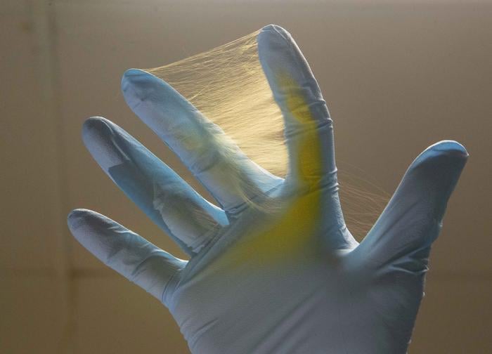
It’s smaller than a thumbnail. A dense, whitish square, barely a millimetre thick, laser-cut from a folded membrane of polymer fibres thinner than human hair. Under a microscope the structure looks almost textile, a woven lattice with fibres crossing and recrossing in layers. Surgeons would place it in the cavity left by tumour removal, where it would sit, slowly releasing its chemical cargo for weeks. The tumour, if it returned at all, would need to fight its way through a three-front pharmaceutical ambush designed specifically to shut down the escape routes it has spent decades learning to use.
This is the NanoMesh, developed by researchers at the University of Cincinnati and Johns Hopkins Medicine, and the results from animal trials are, to put it carefully, rather better than anyone has managed with this tumour in a long time.
Glioblastoma is perhaps the bleakest diagnosis in oncology. The most common primary brain cancer in adults, it kills the vast majority of patients within roughly 18 months of diagnosis. Standard treatment (surgery, followed by radiation and chemotherapy using a drug called temozolomide) has barely shifted that number in two decades. Part of the problem is structural. Glioblastoma is made of heterogeneous cells, meaning no two tumours are quite alike, and within a single tumour there are populations of cells with different vulnerabilities, different mechanisms of resistance, different molecular identities. When you kill one population, another adapts. Treatments knock at one door and the cancer quietly opens another.
Andrew Steckl, a Distinguished Research Professor at UC whose NanoLab pioneered the electrospinning technology behind the mesh, put it more vividly. The cancer, he said, “comes in through the window and when you close the window, it comes through the door. And when you close that, it comes through the chimney.”
Three Drugs, One Mesh, One Problem
The chimney problem is precisely what the NanoMesh was designed to address. Rather than a single drug, the implant carries three, each targeting a different aspect of how glioblastoma survives and spreads. Temozolomide, already the standard chemotherapy for the disease, acts as a DNA-alkylating agent, sabotaging cancer cells’ ability to replicate. The other two, acriflavine and PT2385, are newer additions, both targeting a set of proteins called hypoxia-inducible factors, or HIFs.
HIFs are, in a sense, glioblastoma’s oxygen-crisis management system. As tumours grow quickly they outpace their own blood supply, producing pockets of low oxygen. This triggers HIFs to switch on a cascade of genes that help tumour cells adapt: genes controlling blood vessel growth, genes maintaining stem-cell-like populations that are especially resistant to treatment, genes suppressing the immune response. Acriflavine blocks HIF-1α; PT2385 blocks HIF-2α. Between them, the two drugs attack the adaptation machinery from two angles simultaneously, while temozolomide handles direct cell killing. In cell experiments, the researchers measured an 80% reduction in a gene controlling blood vessel growth, and nearly complete suppression of a metabolic gene that tumour cells under oxygen stress rely on heavily. A marker of cancer stemness fell by a similar amount.
The key question was whether the three drugs would actually cooperate, or simply get in each other’s way. Pharmaceutical combinations can be antagonistic (the combination is actively worse than either drug alone), additive (effects simply sum), or synergistic (the combination outperforms the arithmetic). In trials across several glioblastoma cell lines, including patient-derived tumour samples, the three-drug combination produced synergistic effects in every case. Two-drug combinations gave mixed results depending on the cell line; the third drug was apparently necessary to tip all of them into synergy.
“Unfortunately, cancers know how to pivot to evade therapeutic treatment,” said Betty Tyler, a professor of neurosurgery at Johns Hopkins who worked on the study. “So we’re approaching treatment multidimensionally.”
Getting the Drugs to Stay Put
Identifying a synergistic combination is one thing. Delivering it to a brain tumour, in the right sequence, in useful quantities, without poisoning the surrounding tissue, is something else entirely. This is where the engineering becomes as important as the pharmacology. The blood-brain barrier, a tight cellular seal protecting the brain from circulating toxins, also blocks most systemically administered chemotherapy drugs. The NanoMesh sidesteps this problem by not entering the bloodstream at all. It’s implanted directly, and the same barrier that would have stopped drugs reaching the tumour now works the other way, keeping the high local drug concentrations from leaking into the rest of the body.
The fibres themselves are coaxial: each one has a core carrying the drug payload and an outer sheath of biodegradable polymer. The sheath controls how quickly the core is exposed to fluid and, therefore, how quickly the drug is released. By adjusting the material composition and the fibre dimensions, the team can tune release kinetics independently for each drug. In practice this means temozolomide and acriflavine deliver a large initial dose within the first day or two, providing immediate toxic pressure on the tumour, followed by a slower trickle over weeks. PT2385, the HIF-2α inhibitor, shows a more sustained release profile extending beyond nine weeks. The combination of fast and slow release isn’t accidental: the tumour’s stem-like cells, which are slower to respond to acute chemical injury, need prolonged exposure to an inhibitor like PT2385 to lose their resilience. “Our NanoMesh system was designed to solve these issues by enabling localized long-term delivery of multiple synergistic drugs directly at the tumor site after surgery,” said Daewoo Han, the lead author and an assistant professor in UC’s College of Engineering.
In mouse trials, untreated animals with glioblastoma died within about 19 days. Those receiving the three-drug NanoMesh showed a median survival improvement of more than 50 days. More strikingly, 40% of treated animals were still alive at the 120-day end of the experiment, a plateau the researchers describe as long-term survival. Bioluminescence imaging, which tracks the tumour cells using light-emitting markers, showed the tumour burden remained essentially flat in treated animals over the critical first two weeks while control animals’ tumours expanded rapidly until they died. In a second, more aggressive cancer model, single-drug implants of any of the three compounds individually produced no survival benefit whatsoever. The combination was necessary.
There are caveats. The fast initial release of the hydrophilic drugs is a concern; roughly 70% of the acriflavine and temozolomide exits the fibres within the first 24 hours, which could in principle create a spike of local toxicity. The team are now working on a trilayered fibre design that adds a hydrophobic intermediate barrier between the drug core and the outer sheath, slowing that early burst. And mice are not people. The tumour models used, while well-validated and genuinely aggressive, do not replicate the full complexity of human glioblastoma or the variability of human immune responses. A long road of safety and efficacy testing lies between these results and a clinical device.
Tyler was measured about what existing treatments have achieved. “Current therapies have increased patient survival and given them more birthdays,” she said. “But we’re still working on improving options.”
What the NanoMesh represents, for now, is a fairly coherent answer to a specific structural problem in cancer therapy: tumours that mutate around single-drug approaches. Whether the same platform could carry different drug combinations for other cancers is something the UC team has started thinking about. The electrospinning process is, in principle, drug-agnostic; the fibre architecture can be adapted for different molecular payloads. Glioblastoma is the test case. If it works there, of all places, it might work anywhere.
https://doi.org/10.1021/acsbiomaterials.5c01482
Frequently Asked Questions
What makes the NanoMesh different from the Gliadel wafer already used in brain cancer surgery?
The Gliadel wafer carries a single drug, carmustine, and releases it over a relatively short period, which allows heterogeneous tumour cells to develop resistance. The NanoMesh delivers three drugs simultaneously from a layered fibre structure that provides both rapid initial dosing and sustained release over weeks to months. The triple combination was specifically chosen because it produced synergistic effects across multiple glioblastoma cell lines, something single-drug approaches cannot achieve.
Why does glioblastoma become resistant to chemotherapy so readily?
Glioblastoma is made up of genetically diverse cells, so even when one drug kills the majority of tumour cells, populations with different vulnerabilities survive and continue growing. The tumour also contains stem-like cells that are especially resistant to standard chemotherapy and help the cancer rebuild after treatment. Two of the three drugs in the NanoMesh directly target the molecular machinery these stem cells use to survive under stress.
Could the NanoMesh approach be used for other types of cancer?
In principle, yes. The electrospinning process that produces the fibres can incorporate different drug payloads, and the release kinetics can be tuned for different compounds. The researchers describe the platform as broadly applicable to difficult-to-treat cancers. However, every new drug combination would require its own safety and efficacy testing, so translation to other cancer types would involve substantial additional research.
What does the 40% long-term survival figure actually mean at this stage?
It means 40% of the mice treated with the three-drug NanoMesh in the primary animal model were alive at the 120-day endpoint of the experiment, compared with zero in the untreated group. These are encouraging results, but mouse tumour models do not perfectly replicate human glioblastoma, and the path to human trials involves further toxicity testing, dose optimisation, and larger preclinical studies. The figure is meaningful as proof of concept, not as a prediction of human outcomes.