

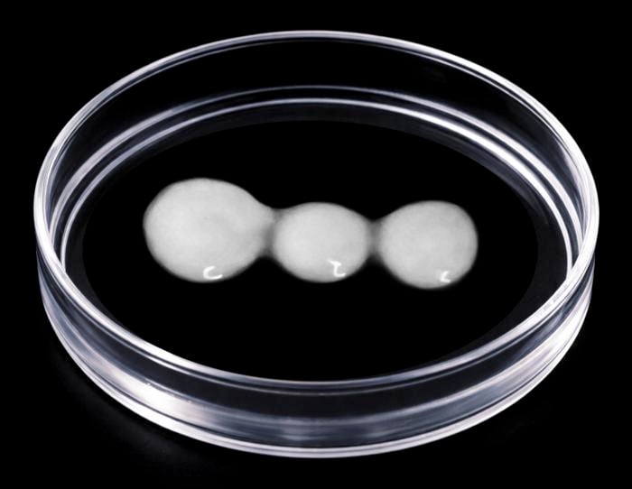
The muscle cluster flinched. Not a twitch caused by direct stimulation, but a contraction driven by signals travelling along axons that had grown, over weeks, from a miniature brain across a gap and into a miniature spinal cord. Nobody had seen this in a dish before, at least not like this, with the cortex and spine kept separate, connected only by the nerve fibers threading between them. George Gibbons, the Cambridge PhD student who had spent months coaxing these tissues into existence, watched on a microscope feed as the myosphere pulsed in response to electrical prompting of the cortical side. The circuit worked. Now they could break it, deliberately, and see what happened next.
What they found has implications for some of the most intractable injuries in medicine. Paralysis after spinal cord damage is usually permanent because the axons that carry movement signals from brain to spine almost never regrow. That has been understood for decades. What has been less clear is why they stop being able to grow in the first place, and whether that inability is truly fixed or simply a developmental state that could, in principle, be reversed.
When the Wiring Stops Repairing Itself
The Cambridge team, led by Dr András Lakatos at the Department of Clinical Neurosciences, published their results in Cell Reports this week. The model they built is called a corticospinal connectoid: cortical organoid slices grown at an air-liquid interface on one side, spinal organoid slices on the other, roughly two millimetres of bridging gel between them. Axons from the cortical tissue grew into the spinal tissue, formed functional synapses with motor neurons and interneurons, and eventually triggered those muscle contractions. The system was sustained for more than a year, long enough to ask a question that has not previously been possible to ask using human tissue: at what point in development does the central nervous system lose the ability to repair its own axons?
The answer, it turns out, is surprisingly precise. And, importantly, not quite as final as it looked.
Gibbons and colleagues injured neurons taken from organoids at different developmental stages: roughly corresponding to 75, 100, 150 and 290 days in vitro, mapping onto a window spanning the second trimester of pregnancy through to early postnatal life. The younger neurons, those pulled from organoids at around day 75 to 100, regrew their fibres reasonably well. The older ones did not. “Neurons taken from less mature organoids regrew long fibres after injury, but those from more mature organoids showed a sharp drop in their ability to regrow,” Gibbons said. “In other words, poor regeneration is built into human neurons as they mature in the central nervous system.”
That phrase, “built into,” is doing a lot of work. It suggests the failure of axon regeneration is not just a consequence of the injured environment (the scar tissue, the inflammatory signals, the inhibitory molecules that accumulate at injury sites) but a property of the neurons themselves, encoded during development. Which raises the obvious question: encoded how?
A Switch Buried in the Genome
The team turned to single-cell transcriptomics to find out. Using RNA sequencing across the organoid time points, they identified a network of genes in the deep-layer cortical projection neurons that shifts significantly between the 150 and 290-day stage, precisely when axon regrowth ability drops off. The network has about 20 core regulatory nodes, including genes involved in cytoskeletal structure, protein transport, RNA metabolism, energy supply, and synaptic adhesion. Several of these connect strongly to PTEN, a gene already known to restrict axon extension in mice. When they inhibited PTEN in their model using a compound called VO-Ophic, growth cone movement at injury sites increased nearly threefold within about an hour of treatment.
So the network could be turned back, or partially back, toward a more regenerative state. That confirmed the platform was working as a target-discovery tool. The next step was finding something that might actually be usable.
The team ran the full gene network through several FDA-approved compound databases and generated a list of 323 candidates. Six were selected for direct testing in cortical neuron cultures. Lynestrenol, a synthetic progesterone receptor agonist that has been licensed since the 1960s as a treatment for endometriosis and as a contraceptive, came out on top. When applied to mature cortical neurons following axon injury in microfluidic chambers, it produced roughly a twofold increase in axon length after five days. The drug, it appears, does something to the transcriptional state of the mature neuron that allows its growth machinery to partially reactivate. The precise downstream mechanism is not yet worked out.
Lakatos was measured about what this means. “Lynestrenol itself may not be the answer to spinal cord repair, but it shows us that, in principle, it should be possible to directly target human neurons and regenerate their axons,” he said. “Although we still need to show that this strategy will also help to re-establish appropriate connections between the brain and spinal cord cells, this gives us hope that one day we may be able to treat conditions previously thought untreatable.”
Why Human Tissue Changes Everything
A fair question at this point is why any of this required an elaborate in-vitro brain-spinal cord circuit. The answer has to do with species. Much of what neuroscience knows about axon regeneration comes from rodents, and rodent neurons behave differently from human ones in ways that matter. Mouse neurons, for instance, show a developmental restriction of axon growth, but the timing, the transcriptional mechanisms and the drug responses don’t necessarily translate. Lakatos pointed out that the corticospinal tract in primates even wires differently from that of rodents, with cortical neurons projecting directly onto motor neurons in a way that is essentially unique to certain primates including humans. Without a human system, you can test interventions that look promising in mice and see them fail in patients, because you were never really studying the same thing.
There are real limits to what the connectoid model can currently tell you. It lacks immune cells, blood vessels, and connective tissue, all of which contribute to the non-permissive environment that blocks repair at real injury sites in adults. And it was built from a single human embryonic stem cell line, which means individual genetic variation is not yet part of the picture. Whether the drug effects seen here survive contact with those complicating factors remains genuinely unknown.
But the model also provides something the field has not previously had: a human developmental timeline for a failure that, for most of recorded neuroscience, has been treated as simply fixed. The cutoff at around 150 days in vitro corresponds to mid-pregnancy. That is when the switch appears to close. And it is, apparently, still a switch, not a one-way door. That distinction, modest as it sounds, is where the hope lives.
https://doi.org/10.1016/j.celrep.2026.117399
Frequently Asked Questions
What is a corticospinal connectoid and why does it matter?
A corticospinal connectoid is a lab-grown model that mimics how the brain’s motor cortex connects to the spinal cord. Cambridge researchers built it by growing miniature versions of both tissues separately and allowing nerve fibres from the brain tissue to bridge the gap and form working connections with the spinal tissue. The key advantage is that the two regions remain spatially distinct but functionally linked, which means researchers can study the cortical neurons specifically without the results being muddied by direct contact with spinal cells. It is the most faithful human model of this neural circuit built to date, and it can be kept alive for more than a year.
Why can’t the spinal cord repair itself after injury?
Two things work against repair. One is the external environment at the injury site: scar tissue, inflammatory signals, and inhibitory molecules that make it physically difficult for axons to grow through. The other, and the one this study focuses on, is internal to the neurons themselves. As the brain’s motor neurons mature during fetal and early postnatal development, they progressively lose the molecular machinery needed to regrow axons. This study found that this internal shutdown happens around the mid-trimester of pregnancy and is encoded by a specific network of genes. That internal block, not just the external environment, is a major reason why paralysis after spinal cord injury tends to be permanent.
How was lynestrenol identified as a potential treatment?
The researchers first identified the gene network responsible for switching off axon growth during development. They then ran that network against databases of FDA-approved compounds to find drugs whose known molecular targets overlapped with the network. Around 323 candidates emerged; six were tested in cortical neuron cultures, and lynestrenol produced the biggest improvement in axon regrowth after injury. The drug is a synthetic hormone that has been used for decades as a contraceptive and for managing endometriosis, and it has a relatively favourable side-effect profile. Why it activates the axon growth machinery is not yet fully understood.
Does this mean paralysis could be reversed?
Not yet, and probably not directly from this work alone. Lynestrenol showed promise in cell culture experiments, but the model used here lacks the immune cells, scar tissue, and blood vessels that also block regeneration in real spinal cord injuries. Any drug that successfully restores a neuron’s ability to grow would also need those regrowing axons to find their correct targets and form functional connections, which is a separate challenge. The researchers themselves are cautious: this study establishes that the internal growth block can be reversed in principle, and identifies a starting point for drug development, but substantial further work is needed before human trials become realistic.
What is the advantage of using human organoids over animal models?
Rodent neurons, which have provided most of what neuroscience knows about axon regeneration, behave differently from human neurons in ways that often matter clinically. The timing of developmental changes, the transcriptional mechanisms involved, and the response to drugs can all differ between species. Several treatments that worked in mice have not translated to patients, partly because the biology was not the same to begin with. Human organoids grown from stem cells replicate human developmental timelines and gene expression patterns, which makes findings more likely to apply to actual patients. They also sidestep some of the ethical issues around animal research, though they have their own limitations, including the absence of immune and vascular components.