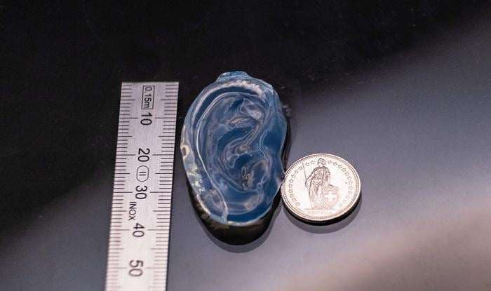
The ear appears all at once. A glass vial of clear gel, a stage rotating beneath a 150-milliwatt blue laser diode, a hologram flickering through 1,440 frames per second, and after 2 minutes and 12 seconds, suspended in the middle of the cylinder, a life-size human ear. No layers. No support scaffolding. Just a complete, three-dimensional object that has materialized inside the gel like a photograph developing in reverse, only sculptural.
That, roughly, is what tomographic volumetric additive manufacturing now looks like in the basement of a Swiss engineering school. And the basement in question, the Laboratory of Applied Photonic Devices (LAPD) at the Ecole Polytechnique Fédérale de Lausanne, has just published results that bring the technique closer to something we will probably all care about eventually: bioprinted implants made from living cells.
The trick, the one that makes this whole approach feasible, is that you do not build the object up. You shine carefully patterned light through a rotating vial of photosensitive resin, and wherever enough light arrives, the resin hardens. The geometry is encoded in the light itself, in the way successive holographic projections, taken together, deposit a precise three-dimensional dose. Think of it as a sort of inverse CT scan: instead of taking pictures from many angles to reconstruct an object, you fire patterns from many angles to construct one.
The catch, until now, has been efficiency. Conventional volumetric printers use a digital micromirror device, an array of tiny mirrors that toggle on and off, which throws away most of the laser’s power. Only a few percent of the light actually makes it into the resin.
What Christophe Moser, who heads the LAPD, and his PhD student Maria Alvarez-Castaño have done is swap that lossy modulator for a different beast entirely: a phase light modulator, or PLM, built around a chip whose pixels are micromirrors that move up and down in tiny piston motions rather than tilting on and off. Each mirror imparts a phase delay on the light hitting it, which lets the team encode the projection as a hologram rather than a binary image. The upshot, reported this month in Light: Science & Applications, is a 70-fold jump in light efficiency over the amplitude-based approach, and about twice the efficiency of a previous holographic version the same group demonstrated last year.
“Our method’s demonstrated efficiency and precision finally makes it possible to bioprint tissue-like structures at near-clinical scale,” says Moser.
Light That Doesn’t Mind the Cells
So what does that actually buy you? Quite a lot, as it turns out. The new system runs at up to 1,440 holograms per second, lets the team print millimeter-scale things in seconds and centimeter-scale things in minutes, and, crucially, copes with cloudy stuff. Cells, when you suspend a million of them per millilitre in a hydrogel, scatter light in awkward ways; conventional volumetric printing tends to lose fidelity quickly in such media unless each batch is painstakingly characterized in advance. Alvarez-Castaño and colleagues sidestep that problem by shaping each holographic projection into a Bessel beam, using an axicon phase pattern that produces a slim, low-divergence column of light. Bessel beams have a quirk that physicists sometimes call self-healing: scatter them with an obstacle and they reform downstream. In a vial full of cells, that turns out to be exactly what you want.
To prove the point, the team printed a multiacinar construct, a four-millimetre cube laced with internal cavities meant to mimic the tubuloacinar architecture of the exocrine pancreas, in a gel containing one million human fibroblasts per millilitre. Six days later, under a confocal microscope, the cells were not just alive; they had organized themselves into elongated networks around the cavities. Eight times the volume, twice the cell density, compared with the lab’s earlier work. “We have printed structures substantially larger than those achieved with previous holographic approaches, despite increased light scattering caused by the embedded cells,” Moser notes.
Speckle, though, is the other niggle, and the team has had to wrestle with it. Holograms produced by phase-only modulators are prone to grainy interference patterns, the same speckle you see in a laser pointer dot on the wall, which can leave printed surfaces bumpy and even cause delamination during printing. The fix is delightfully simple in concept and rather fiddly in practice: compute nine slightly different holograms per projection angle, each shifted by roughly half a speckle-grain in a different direction, and play them in sequence so the bright bits and the dark bits average each other out. The resulting prints come out visibly smoother. A DNA double helix produced with and without the technique sit side by side in the paper, and the difference is obvious to the eye.
From Fusilli to Implant
To stress-test the light engine, the LAPD team printed a small menagerie of objects in commercial acrylate resin. A 32-second fusilli at 18 milliwatts. A Stanford Bunny in just over a minute. A pair of DNA helices, the smaller one resolving cross-bars only tens of micrometres across, the smallest positive feature they have managed. Then they swapped to the gels: GelMA loaded with fibroblasts for the pancreas-like construct, and gelatin thiol-norbornene, a more reactive material that copes better with oxygen, for the ear. For comparison, an acrylate version of the same ear printed at the same scale took nearly eight minutes; conventional tomographic printers typically need lasers around 6 watts to manage objects of comparable size. The PLM-driven system used a fortieth of that.
None of which means tissue-grade ears are walking out of EPFL next year. The group is candid about the remaining limits. Features below 50 micrometres still get gnawed at by oxygen diffusion, which inhibits the polymerization chemistry; the holographic projections themselves carry phase-quantization errors from the modulator’s 16 discrete levels; and machine-learning approaches to refining the projections are flagged as a next step rather than a current capability.
Still, there is a reason the ear-shaped milestone matters. Reconstructive surgery for missing or damaged ears currently relies on harvested cartilage or moulded silicone, neither ideal. A bioprinted construct seeded with a patient’s own cells, fabricated in minutes from a low-power laser diode that costs a fraction of the gear conventional volumetric printers require, would be a different proposition entirely. Alvarez-Castaño puts it in the press release: the approach brings volumetric printing closer to real-scale implants and biologically compatible manufacturing using low-power laser sources. The group already has further papers in preparation, describing how to print directly onto or around existing objects, and how to model the resin chemistry well enough to capture features finer than oxygen will currently allow.
Whatever comes next, the basement at EPFL has a glass vial in it, and inside that vial something complicated keeps appearing all at once.
https://doi.org/10.1038/s41377-026-02331-4
Frequently Asked Questions
How is volumetric 3D printing different from regular 3D printing?
Regular 3D printers build objects layer by layer, depositing material one slice at a time. Volumetric printing solidifies an entire three-dimensional shape at once by shining patterned light through a rotating vial of photosensitive resin, with the geometry encoded in how the light doses overlap from different angles. The result is no layer lines, far faster print times for complex shapes, and the ability to embed delicate things like living cells without mechanical stress.
Why does it matter that the system uses a phase light modulator instead of a regular mirror chip?
The standard chip used in volumetric printing throws away most of the laser’s power because each pixel can only be fully on or fully off. A phase light modulator instead nudges each mirror up and down by tiny amounts, shifting the timing of the light wave so the projection can be shaped as a hologram. That recovers around 70 times more of the laser’s output, which is what makes printing centimetre-scale objects with a cheap 150-milliwatt diode possible.
Could this actually be used to print human tissue for transplants?
Not yet, but the EPFL team has now printed cell-laden structures at near-implant scale, including a life-size human ear in a gelatin gel and a centimetre-scale construct seeded with one million fibroblasts per millilitre. The cells stayed alive and organized into networks over six days, which is a meaningful step. Getting to clinical use will still require working out vascularization, regulatory approval, and finer feature control around 50 micrometres and below.
What’s stopping the technique from making even more detailed prints?
Two main things. The first is oxygen diffusion: oxygen inhibits the chemistry that hardens the resin, and for very small features it nibbles away the edges before they can fully set. The second is that the holographic projections themselves have small imperfections, partly because the modulator only offers 16 discrete phase levels rather than a smooth continuous range. Machine learning and modified resin chemistries are both being explored to address these limits.
How does the system manage to print inside a vial full of cells without the image blurring?
The team shapes each holographic projection into a Bessel beam, a slim column of light created by applying an axicon-style phase pattern to the hologram. Bessel beams have a self-healing property: when they encounter a scattering object, they reform downstream rather than dispersing. That property turns out to be ideal for printing through a hydrogel packed with cells, which would otherwise scatter the light badly.
ScienceBlog.com has no paywalls, no sponsored content, and no agenda beyond getting the science right. Every story here is written to inform, not to impress an advertiser or push a point of view.
Good science journalism takes time — reading the papers, checking the claims, finding researchers who can put findings in context. We do that work because we think it matters.
If you find this site useful, consider supporting it with a donation. Even a few dollars a month helps keep the coverage independent and free for everyone.