At rest, without any breath-holding or injections or particular effort, your brain is doing something quite extraordinary. Its blood vessels are constantly micro-adjusting — dilating and constricting in response to tiny fluctuations in blood pressure and carbon dioxide — keeping the roughly 100 billion neurons inside bathed in a precise supply of oxygen. It’s a system so automatic we never notice it. But Amaryllis Tsiknia, a PhD candidate at the University of Southern California’s neuroimaging institute, reckons that when this system starts to falter, the telltale signs might appear years before any memory problems do.
Her team has now found evidence to back that hunch up. In a study published this month in Alzheimer’s & Dementia, Tsiknia and colleagues showed that subtle differences in how efficiently the brain regulates its own blood flow and oxygen delivery are closely tied to two hallmark features of Alzheimer’s disease: amyloid plaque buildup and shrinkage of the hippocampus, the seahorse-shaped structure deep in the temporal lobe that’s essential for forming new memories.
The measurements involved no MRI scanners, no radioactive tracers, no demanding protocols. Participants simply lay still for eight minutes while the team monitored them with two noninvasive tools. Transcranial Doppler ultrasound tracked blood velocity in a major brain artery by bouncing sound waves through the skull; near-infrared spectroscopy, working like a pulse oximeter but positioned over the forehead, measured how much oxygen was reaching the cortex. From these continuous signals, an advanced mathematical model extracted five distinct indices — each capturing a slightly different aspect of how the vasculature responds to spontaneous pressure and CO2 changes.
Higher values on those indices meant the brain’s vessels were behaving more like those of cognitively healthy adults. Lower values suggested something was off.
“Amyloid and tau are often considered the primary players in Alzheimer’s disease, but blood flow and oxygen delivery are also critical,” says Tsiknia. “Our results show that when the brain’s vascular system functions more like it does in healthy aging, we also see brain features that are linked to better cognitive health.” Participants whose vascular indices fell into the healthier range tended to have less amyloid deposited across their cortex and, separately, larger hippocampal volumes — both well-established markers of lower Alzheimer’s risk, routinely assessed by PET and MRI scans costing thousands of pounds a session.
What’s perhaps most intriguing is that the associations didn’t vanish even after the researchers accounted for age, sex, APOE ε4 status (the main genetic risk factor for the common form of the disease), and cognitive test scores. The vascular signal was doing something independent. Senior author Meredith Braskie, an assistant professor of neurology at the Keck School of Medicine, puts it this way: “These vascular measures are capturing something meaningful about brain health. They appear to align with what we see on MRI and PET scans that are commonly used to study Alzheimer’s disease, providing important information about how vascular health and standard brain measures of Alzheimer’s disease risk may be related.”
The findings slot into a growing body of research — call it the vascular hypothesis of Alzheimer’s — suggesting that the disease isn’t purely a story of amyloid and tau proteins gone wrong. Vascular risk factors like hypertension are associated with greater dementia risk; treating high blood pressure appears to reduce it. The proposed mechanisms converge on a common problem: when blood supply to the brain falters chronically, neurons get less oxygen, the blood-brain barrier weakens, and conditions that encourage amyloid accumulation kick in. Animal studies have gone further, showing the relationship is probably bidirectional — amyloid plaques themselves impair vascular regulation, which in turn accelerates further plaque deposition. A vicious cycle, in other words.
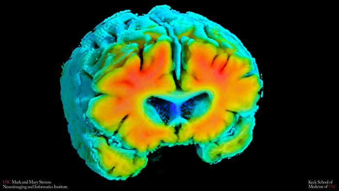
The hippocampus may be especially susceptible to all this, which is perhaps why memory is usually the first thing to go. The microvasculature serving hippocampal tissue is architecturally distinct from that in the neocortex — its capillaries are sparser, neurovascular coupling weaker — making it rather more vulnerable to even subtle reductions in perfusion.
But the study brings a practical angle too. Arthur Toga, director of the Stevens Neuroimaging and Informatics Institute and one of the paper’s co-authors, notes that “understanding how blood flow and oxygen regulation interact with amyloid and brain structure opens new doors for early detection and potentially prevention.” The tools required here — a handheld ultrasound probe and a spectroscopy headset — are portable, relatively cheap, and don’t require patients to hold their breath on command or withstand the claustrophobic tube of an MRI scanner. That matters a great deal for widespread screening, or for older patients who cannot tolerate more demanding procedures.
There are caveats, naturally. The study was cross-sectional, meaning it captured a snapshot rather than tracking people over time. It therefore can’t settle the chicken-and-egg question: does poor vascular regulation cause the Alzheimer’s-related changes observed, or does early Alzheimer’s pathology itself damage the vasculature? Both seem plausible. The sample was also mostly non-Hispanic white adults with relatively low cardiovascular risk — a limitation the authors are candid about, given that dementia disproportionately affects Black and Hispanic populations in the United States.
Longitudinal data collection is already underway across the three study sites (USC, the University of Kansas, and UT Southwestern). “If we can track these signals over time,” Tsiknia says, “we may be able to identify people at higher risk earlier and test whether improving vascular health can slow or reduce Alzheimer’s-related brain changes.” That possibility — that something as manageable as blood pressure treatment or exercise might move these indices in the right direction — gives the work an urgency beyond the immediate findings.
Whether or not the vascular angle eventually reshapes how we screen for Alzheimer’s risk, the study adds weight to a view that’s been gaining ground for years: that the brain’s plumbing deserves at least as much attention as the proteins clogging it up.
Study link: https://alz-journals.onlinelibrary.wiley.com/doi/10.1002/alz.71146
ScienceBlog.com has no paywalls, no sponsored content, and no agenda beyond getting the science right. Every story here is written to inform, not to impress an advertiser or push a point of view.
Good science journalism takes time — reading the papers, checking the claims, finding researchers who can put findings in context. We do that work because we think it matters.
If you find this site useful, consider supporting it with a donation. Even a few dollars a month helps keep the coverage independent and free for everyone.