Somewhere in the mineral lattice of your skeleton, calcium and phosphate ions are locked into a crystalline structure called hydroxyapatite. It is, in a sense, what you are made of: the same compound accounts for roughly 70 percent of bone’s dry weight, giving it a rigidity that resists compression and a chemistry that living cells recognise instinctively. For decades, surgeons reaching for bone graft material had to look elsewhere: to cadaver donors, to a patient’s own hip or tibia, to synthetic substitutes that the body would accept, reluctantly, as an approximation. Now a team in Finland think they have found a better answer. Use the real thing. Just print it.
More than 2 million bone grafts are performed worldwide every year, making it the second most common tissue transplantation procedure after blood transfusions. Demand is climbing as populations age.
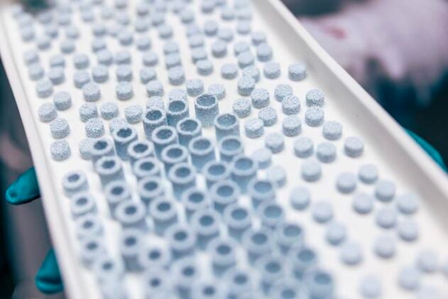
Antonia Ressler, a postdoctoral fellow at Tampere University’s Institute for Advanced Study, has spent four years working out how to build bone-mimicking ceramic scaffolds using a 3D printing method called vat photopolymerisation. The technique works by exposing a ceramic slurry, layer by layer, to precisely controlled ultraviolet light, hardening the material into complex three-dimensional shapes with internal architectures that a conventional mould could never produce. “By using the same material that nature uses and shaping it through ceramic 3D printing, the implants can be precisely tailored to match a patient’s individual bone defect, without relying on drugs or growth factors that may cause side effects,” Ressler says. It’s a quieter promise than most regenerative medicine makes, and perhaps more credible for that.
Getting the Architecture Right
The hard part is not the chemistry. Hydroxyapatite has been used in bone repair for years, and its biocompatibility is well established. The hard part is the geometry. Natural bone is a hierarchical structure: dense cortical shell, spongy trabecular interior, pores running at multiple scales to carry blood vessels and cells and nutrients. Replicate the chemistry but not the architecture, and you get a material that sits inert in the defect rather than integrating with it.
Ressler and her collaborators designed four scaffold variants with different porosities and pore sizes, then put them through a battery of biological tests using human bone marrow stem cells and osteoclasts. The winner, by some margin, had pores averaging around 400 micrometres and a porosity of roughly 45 percent, a structure that would sit comfortably in the middle range of natural trabecular bone. “This architecture achieved a crucial balance between strength and biological performance, allowing bone-forming cells to enter the material, interact with one another, and successfully begin forming new bone tissue,” she says. Stem cells seeded onto these scaffolds produced collagen type I (the structural protein that bone mineralises around) and osteocalcin, a late marker of bone-forming cell differentiation. Signs, in other words, that the cells were doing what bone cells do.
The osteoclasts are perhaps the more interesting part of the story. Bone is not a static material; it is continuously remodelled, old tissue eaten away by osteoclasts while osteoblasts deposit new matrix in its place. For a synthetic scaffold to work properly long-term, it cannot just sit there being colonised. It has to gradually yield, to be resorbed as the patient’s own bone grows through. The scaffolds based on pure hydroxyapatite allowed osteoclasts to function normally, leaving visible resorption trails in scanning electron microscopy images. Which is exactly what you want.
When Chemistry Turns Against You
There is a complication, and it runs to the heart of what makes biomaterial design so genuinely difficult. Natural bone is not made of pure hydroxyapatite. The mineral in your skeleton contains trace quantities of strontium, magnesium, zinc, and other ions substituted into the hydroxyapatite lattice, each playing some role in the bone formation process. Magnesium, for instance, acts as a growth-factor-like signal in early osteogenesis; zinc supports the enzyme activity needed for matrix mineralisation. Ressler’s team tried to replicate this chemistry, loading their scaffolds with the same trace elements. In principle, these ion-substituted variants should have been superior. They were not.
The problem was heat. Ceramic 3D printing requires sintering, which means firing the printed structure at high temperatures to burn off the organic binder and fuse the ceramic particles. But when strontium, magnesium, and zinc are present in the hydroxyapatite lattice, those temperatures trigger a phase transformation: the hydroxyapatite partially converts to a different calcium phosphate compound called beta-tricalcium phosphate. And that transformed surface, it turns out, is hostile to cells. “We found that the high temperatures required during processing can alter the surface of the material in ways that make it more difficult for human cells to attach,” Ressler says. “Our finding highlights that not only the composition, but also the surface properties of biomaterials are critical for successful bone regeneration.”
The substituted scaffolds had a more negative surface charge and were markedly more hydrophobic than the pure hydroxyapatite versions. Cells prefer to land on wet, moderately charged surfaces; present them with something that repels water and carries excess negative charge, and attachment drops sharply. Osteoclasts, tested on the same material, barely differentiated at all. The ion enrichment that should have improved biological performance had, via a processing detour, undermined the basic requirement for cells to stick to the implant in the first place.
A Decade Away, Perhaps
It is a genuinely useful failure. The ceramic printing approach is still new enough that understanding exactly where the trade-offs lie matters enormously, and Ressler’s team has laid out one of the clearest maps yet of how processing parameters, microstructure, and surface chemistry interact. The mechanical properties of the best scaffolds sit within the lower range reported for trabecular bone, adequate for non-load-bearing applications, not yet suitable for a femur. Sintering at lower temperatures preserves bioactivity but limits densification; sintering at higher temperatures gives you mechanical strength but kills cell attachment. The team settled on 1000 degrees Celsius as the best available compromise, but the constraint is real.
What the work does establish convincingly is that ceramic vat photopolymerisation can produce scaffolds with genuinely bone-like internal architecture, and that the biology responds to that architecture in the way you would hope. Osteocalcin expression. Collagen networks forming between cells. Osteoclastic resorption trails. The cellular machinery for bone regeneration, running on an artificial substrate. “This technology allows implants to be designed for individual needs: no more ‘one size fits all’ solutions,” Ressler says. “We believe these types of implants could be used in routine bone regeneration treatments within the next decade.”
That is a specific claim from someone who knows where the obstacles are. The next phase of work, an ongoing project called GlassBoneS, will focus on composite approaches combining ceramic and polymer components, which might let the team sidestep sintering altogether, and with it the surface chemistry problem that has so far blocked the trace-element route. Whether the ions themselves can then do what they’re supposed to do, once freed from the constraint of high-temperature processing, remains the open question. Your skeleton has been answering it for millions of years.
The study was published in Materials Today Bio: doi.org/10.1016/j.mtbio.2026.103074
Frequently Asked Questions
Why can’t surgeons just use the patient’s own bone for grafts?
They often do, and donor bone from the patient’s own body (called autograft) is still considered the gold standard. The problem is supply: harvesting bone from a second site means a second operation, with its own recovery time, risk of nerve damage, and blood loss. For large defects, there simply may not be enough bone available from the donor site. As the global population ages and demand for bone repairs increases, finding scalable alternatives that don’t depend on limited human tissue is becoming increasingly urgent.
What does “3D-printed bone” actually mean? Is it real bone?
Not quite. The scaffolds are made of hydroxyapatite, the same mineral that forms natural bone, but they contain no living cells when they’re printed. The idea is that the scaffold acts as a template: surgeons implant it into the defect, and the patient’s own bone-forming cells gradually colonise the structure, produce collagen, and eventually replace the scaffold with genuine bone tissue as it’s slowly resorbed. The printed structure is the starting frame, not the finished product.
If the material is identical to bone mineral, why has it taken so long to print it effectively?
The chemistry is only part of the challenge. Natural bone has an extraordinarily complex internal architecture, with interconnected pores at multiple scales that allow cells, blood vessels, and nutrients to penetrate deep into the tissue. Earlier manufacturing techniques couldn’t reproduce that geometry precisely enough. Ceramic vat photopolymerisation, a form of 3D printing using light-cured ceramic slurry, is one of the first methods capable of producing scaffolds with the intricate internal pore networks that bone-forming cells actually need.
Why did adding trace elements like zinc and magnesium make the scaffolds worse, not better?
The ions themselves aren’t the problem : zinc and magnesium genuinely do play important roles in bone formation. The issue is heat. Firing the printed scaffold at high temperatures (necessary to burn off the organic binder and harden the ceramic) causes a chemical phase change when those ions are present, converting some of the hydroxyapatite into a related compound called beta-tricalcium phosphate. That transformed surface turns out to be less attractive to cells, because it’s more hydrophobic and carries a more negative surface charge. The team is now exploring composite approaches that might avoid high-temperature sintering altogether, which could finally unlock the potential of the ion-enriched formulations.
Could this technology help with osteoporosis-related fractures?
Potentially, though not in the way you might expect. Osteoporosis weakens bone systemically, which printing can’t address directly. But bone defects resulting from osteoporotic fractures, where a section of damaged or dead bone needs to be replaced, are precisely the kind of application these scaffolds are designed for. The current versions aren’t strong enough for major load-bearing sites like the hip, but the team’s roadmap points toward composite scaffolds with improved mechanical properties, which could eventually extend the technology into those higher-demand applications.
ScienceBlog.com has no paywalls, no sponsored content, and no agenda beyond getting the science right. Every story here is written to inform, not to impress an advertiser or push a point of view.
Good science journalism takes time — reading the papers, checking the claims, finding researchers who can put findings in context. We do that work because we think it matters.
If you find this site useful, consider supporting it with a donation. Even a few dollars a month helps keep the coverage independent and free for everyone.