In a lab at the University of Tokyo, a new kind of microscope is collapsing two worlds of detail into a single view, capturing both the broad contours of a living cell and the restless jitter of nanoparticles inside it in one shot.
By unifying two powerful but previously separate optical techniques, the system can detect signals across an intensity range about fourteen times wider than conventional microscopes, and it does so without adding any fluorescent labels or dyes. The work, reported in Nature Communications, suggests a path toward gentler, longer-term imaging of cells for basic biology and for quality control in pharmaceutical and biotechnology applications.
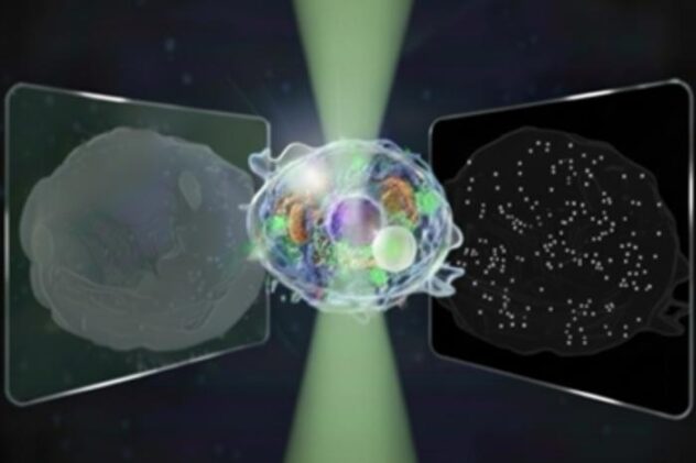
For decades, cutting edge microscopy has been defined by tradeoffs. Quantitative phase microscopy, which measures forward scattered light, excels at mapping whole-cell structure and microscale features such as nuclei and lipid droplets. Interferometric scattering microscopy, which measures backward scattered light, is exquisitely sensitive to nanoscale objects such as single proteins or 50 nanometer beads, and can track their rapid motion. But no single instrument could capture both regimes at once, which meant scientists had to choose between a panoramic view of the cell or a zoomed-in look at the tiniest players that drive its behavior.
Marrying Two Ways Of Seeing Light
The team led by Kohki Horie and colleagues set out to break that compromise by building what they call bidirectional quantitative scattering microscopy, or BiQSM. The system uses off-axis digital holography with bidirectional illumination to record forward and backward scattered light simultaneously on a single image sensor. In a single hologram, forward scattering encodes microscale structure, from overall cell shape to micrometer scale lipid droplets, while backward scattering picks up the faint signatures of rapidly moving nanoscale objects such as small intracellular particles or even features at the cell membrane. By defining a unified scattering-field amplitude for both directions and carefully managing optical shot noise, BiQSM extends the dynamic range of single-frame measurements by about a factor of 14 compared with standard quantitative phase microscopy, enough to visualize everything from whole-cell morphology to 30 nanometer silica beads in one framework.
“Our biggest challenge,” Toda, another first author, explains, “was cleanly separating two kinds of signals from a single image while keeping noise low and avoiding mixing between them.”
Watching Cells Approach Death In Real Time
To prove that this bidirectional approach could do more than just resolve test particles, the researchers turned to one of the most consequential processes in biology, the slow dying of a cell. They used BiQSM to follow living COS7 cells over nearly an hour, acquiring thousands of frames at 500 frames per second and then decomposing each pixel’s motion into low frequency and high frequency components. In the forward scattering images, the method revealed how microscale lipid droplets moved more vigorously at one stage and then slowed as the cell approached death, all while the cell’s total dry mass remained roughly constant. In the backward scattering channel, the team saw distinct changes in the dynamics of nanoscale structures in particle-poor cytoplasmic and nuclear regions, including a drop in slow fluctuations and a surge in fast ones that appeared to synchronize with the behavior of larger droplets elsewhere in the cell.
“We plan to study even smaller particles,” Toda says, already thinking about future research, “such as exosomes and viruses, and to estimate their size and refractive index in different samples. We also want to reveal how living cells move toward death by controlling their state and double-checking our results with other techniques.”
Because BiQSM is label free and relies only on how cells scatter light, it can in principle be applied for long-term monitoring without perturbing the sample, and it can estimate both the size and refractive index of particles directly from the combined forward and backward signals. The authors argue that this opens a route to quantitative characterization of biological nanoparticles in the 100 nanometer to 1 micrometer range, including intracellular vesicles, synthetic drug carriers, and viruses, once background signals from thick, dynamic cell structures are further suppressed. They also suggest that the same bidirectional strategy could be extended to three dimensional imaging, to chemical contrast by pairing with infrared or Raman modalities, and to multimodal systems that add fluorescence. For now, the “great unified microscope” they have built offers a rare thing in optics, a way to see both the forest and the trees inside a living cell, at the very moment its internal life begins to flicker and fade.
Nature Communications: 10.1038/s41467-025-65570-w
ScienceBlog.com has no paywalls, no sponsored content, and no agenda beyond getting the science right. Every story here is written to inform, not to impress an advertiser or push a point of view.
Good science journalism takes time — reading the papers, checking the claims, finding researchers who can put findings in context. We do that work because we think it matters.
If you find this site useful, consider supporting it with a donation. Even a few dollars a month helps keep the coverage independent and free for everyone.