Two patients walk into a psychiatrist’s office with identical depression symptoms—severe low mood, fatigue, and concentration problems. Their brain scans, however, tell completely different stories.
New research from Washington University reveals that depression’s complexity runs far deeper than clinical symptoms suggest, with multiple brain patterns potentially producing the same mental health struggles.
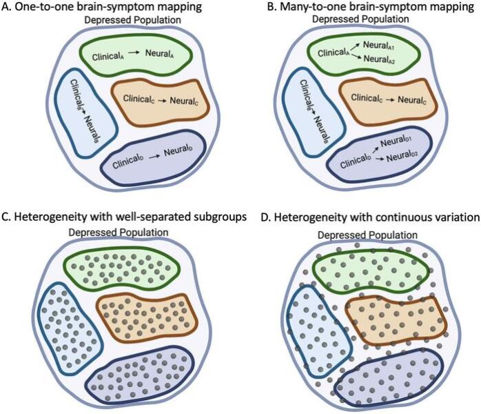
The study, published in Biological Psychiatry, challenges how we understand depression’s neurobiological foundations. Using brain imaging data from over 6,000 people in the UK Biobank, researchers discovered that patients with matching depression symptoms can have distinctly different brain activity patterns—a phenomenon they call “many-to-one brain-symptom mapping.”
Breaking Down Depression’s Building Blocks
“Heterogeneity in depression, i.e., differences between patients with the same diagnosis, has been a topic of interest in our field for a long time,” explains lead investigator Janine D. Bijsterbosch, PhD, from Washington University’s Mallinckrodt Institute of Radiology. The team wanted to understand whether clinical symptoms and brain changes follow simple one-to-one relationships or more complex patterns.
Rather than studying depression as a monolithic condition, researchers isolated specific components: anhedonia (inability to feel pleasure), depressed mood, physical symptoms, chronic episodes, late-onset depression, and acute severe impairment. Each group showed distinct brain signatures when compared to people with mixed symptoms.
The late-onset depression group revealed particularly surprising results. Instead of the expected brain tissue loss, these patients showed preserved or even increased gray matter volume in key regions. This contradicts previous assumptions that brain atrophy drives late-life depression, suggesting instead that larger brain volume might represent a protective mechanism linked to recovery.
Hidden Cognitive Divisions
The most striking discovery emerged when researchers looked deeper into patients with acute depression. Despite having identical symptom profiles, brain imaging revealed two distinct subgroups with dramatically different cognitive abilities.
“Our research also showed that more than one brain profile gave rise to the same clinical presentation in patients with acute depression, providing concrete evidence of many-to-one brain-symptom mapping for the first time,” notes co-investigator Yvette I. Sheline, MD, from the University of Pennsylvania.
One subgroup maintained normal cognitive function despite severe depression, while the other showed significant cognitive impairment. Standard depression questionnaires couldn’t distinguish between these groups—only brain scans revealed the difference.
Key Findings That Reshape Depression Understanding:
- Patients grouped by isolated symptoms showed stronger brain abnormalities than those with mixed presentations
- Late-onset depression was linked to preserved brain volume, not expected tissue loss
- Two cognitively distinct subgroups existed within severe depression cases
- Depressed mood specifically connected to brain regions involved in rumination
The Normative Modeling Advantage
The researchers employed a sophisticated technique called normative modeling, which measures how each patient’s brain deviates from healthy population norms while accounting for age, sex, and scanner differences. This approach proved more sensitive than traditional group comparisons, revealing brain patterns that might otherwise remain hidden.
Critically, the study found that when depression symptoms were mixed together in heterogeneous groups—as typically happens in clinical practice—the brain signal became weaker and harder to detect. This explains why previous neuroimaging studies of depression have produced inconsistent results.
Clinical Implications and Future Directions
“Depression is a very heterogeneous medical condition. The inability to accurately subtype patients is a major obstacle to matching individual patients to treatments that are more likely to be effective for them,” comments John Krystal, MD, Editor of Biological Psychiatry.
The cognitive differences found in the acute depression subgroups hint at practical applications. Brain imaging might eventually help predict which patients will experience cognitive problems alongside their mood symptoms, enabling targeted interventions.
However, questions remain. The study focused on older adults (average age 61) from the UK Biobank, limiting generalizability to younger populations and diverse ethnic groups. The researchers also created artificial groups by isolating single symptoms—most real patients experience multiple depression features simultaneously.
Co-investigator Deanna M. Barch, PhD, from Washington University, concludes: “Identifying distinct subtypes of depression that may respond differently to treatment could greatly improve clinical care for patients with depression in the future. However, our findings show that identifying such subtypes of depression will only be achievable by addressing both clinical and neurobiological heterogeneity.”
This research suggests that effective depression treatment might require both symptom assessment and brain imaging—a personalized medicine approach that recognizes the condition’s hidden complexity beneath seemingly similar presentations.
ScienceBlog.com has no paywalls, no sponsored content, and no agenda beyond getting the science right. Every story here is written to inform, not to impress an advertiser or push a point of view.
Good science journalism takes time — reading the papers, checking the claims, finding researchers who can put findings in context. We do that work because we think it matters.
If you find this site useful, consider supporting it with a donation. Even a few dollars a month helps keep the coverage independent and free for everyone.