Picture this: A researcher places a few skin cells between two electrodes, flips a switch, and within minutes knows which cells are young and which have crossed into the twilight of senescence—all without touching them with a single chemical.
It sounds like science fiction, but it’s happening right now in a Tokyo laboratory.
For decades, scientists hunting aged cells in human tissue have been stuck with a Catch-22. The very process of identifying these cellular troublemakers—using fluorescent tags and biochemical markers—changes the cells themselves, like trying to study a wild animal by first putting it in a cage.
Now, researchers at Tokyo Metropolitan University have found an elegant workaround that reads a cell’s age like a fingerprint, using nothing more than alternating electric fields.
The Electric Dance of Aging
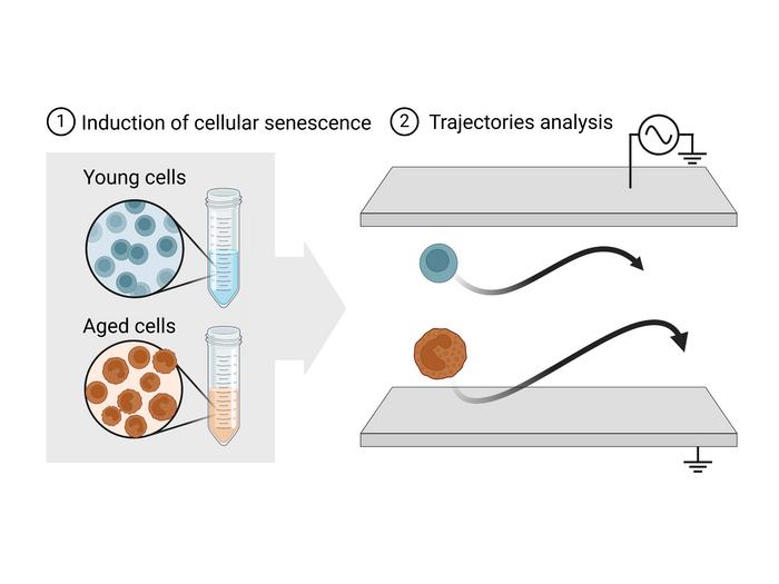
When Assistant Professor Ippei Yagi’s team applies an electric field to human skin cells, something remarkable happens. The cells don’t just sit there—they start moving, migrating back and forth between electrodes in a microscopic waltz.
But here’s where it gets interesting: aged cells dance differently than young ones. As the electrical frequency changes, each cell hits what researchers call a “cutoff frequency”—the moment when its movement pattern shifts dramatically. Think of it as each cell’s unique electrical signature.
“This method, known as frequency-modulated dielectrophoresis (FM-DEP), aims to characterize cell type by measuring this value,” the team explains in their study published in IEEE Sensors Journal.
The difference lies in the cell’s membrane—specifically, changes in fatty lipid molecules that occur as cells age. These molecular shifts alter how the cell responds to electric fields, creating a detectable aging signature.
Why This Matters Beyond the Lab
Consider what’s at stake here. Senescent cells are biology’s equivalent of bitter retirees who won’t leave the workplace—they’ve stopped doing their jobs but stick around causing trouble. These cellular curmudgeons pump out inflammatory compounds linked to arterial hardening, Alzheimer’s disease, and type 2 diabetes.
Understanding how these cells contribute to disease requires studying them in their natural state. But current detection methods create a fundamental problem:
- Chemical labels take hours to attach and can change cell behavior
- Fluorescent markers require complex preparation procedures
- The labeling process itself may trigger cellular stress responses
- Results may reflect the detection method rather than true cell properties
It’s like trying to study how people behave naturally while they’re wearing bright orange jumpsuits—the method influences the outcome.
From Skin Deep to Clinical Promise
Yagi’s team focused on human dermal fibroblasts—the cells that keep our skin strong and help wounds heal. When they compared senescent fibroblasts to their younger counterparts, the electrical signatures were consistently different.
The implications ripple outward quickly. Regenerative medicine researchers, who need to distinguish healthy cells from aged ones for treatments, could benefit from this rapid, non-invasive assessment. Drug developers screening potential anti-aging compounds could test effects on cells without worrying whether their detection method is skewing results.
But perhaps most intriguingly, this electrical window into cellular aging might eventually help doctors assess biological age in patients—moving beyond the crude measure of chronological years to understand how fast someone is actually aging at the cellular level.
The Road Ahead
The researchers are honest about current limitations. They’ve tested only one type of skin cell so far, and the broader applicability remains an open question. Different cell types might have entirely different electrical aging signatures—or the method might not work at all for some tissues.
Still, the elegance of the approach is compelling. In a field where researchers have long struggled with the tools changing the very thing they want to study, sometimes the best solutions are surprisingly simple. Sometimes you don’t need to add anything at all—you just need to listen to what the cells are already saying, in their own electric language.
ScienceBlog.com has no paywalls, no sponsored content, and no agenda beyond getting the science right. Every story here is written to inform, not to impress an advertiser or push a point of view.
Good science journalism takes time — reading the papers, checking the claims, finding researchers who can put findings in context. We do that work because we think it matters.
If you find this site useful, consider supporting it with a donation. Even a few dollars a month helps keep the coverage independent and free for everyone.