Researchers at the Korea Advanced Institute of Science and Technology (KAIST) have developed a sophisticated 3D brain-mimicking platform that can maintain neural activity for 27 days—nearly twice as long as previous systems. The platform achieves six times higher precision than conventional methods while successfully replicating the brain’s complex layered structure.
The research team, led by Professors Je-Kyun Park and Yoonkey Nam from KAIST’s Department of Bio and Brain Engineering, combined three key technologies to overcome a fundamental challenge in neuroscience: creating brain-like structures that are both structurally stable and biologically functional.
Solving the Structure-Function Puzzle
Traditional 3D brain models face a critical trade-off. High-viscosity materials provide structural stability but limit neural growth, while low-viscosity gels that support neuron development are difficult to pattern precisely. The KAIST team solved this by using a “capillary pinning effect” technology.
By printing dilute hydrogels onto stainless steel mesh, the researchers achieved structures with resolution of 500 micrometers or less—six times more precise than conventional methods. The mesh acts like a molecular scaffold, holding the low-viscosity gel in place while allowing neurons to grow naturally.
The platform’s second innovation is a cylindrical 3D printing aligner that ensures printed layers stack without misalignment. This guarantees accurate assembly of multilayer structures and stable integration with microelectrode chips that monitor neural activity.
Dual-Mode Analysis Reveals Neural Conversations
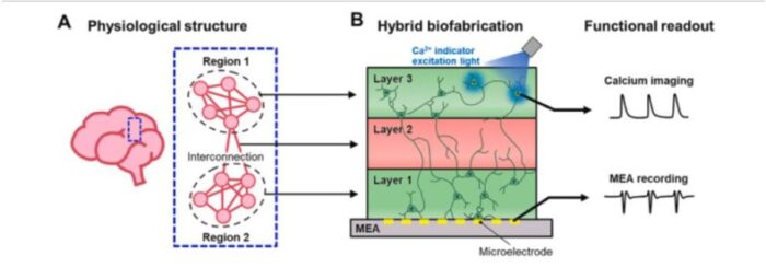
Perhaps most remarkably, the system can simultaneously measure electrical signals from below and observe cell activity with light from above. This dual-mode analysis allows researchers to verify that neurons in different layers are actually communicating—not just sitting adjacent to each other.
In experiments, the team created a three-layered mini-brain with cortical neurons in the upper and lower layers and an empty middle layer designed to allow neural connections. When they applied electrical stimulation, neurons in both layers responded simultaneously. When they blocked synaptic transmission with drugs, the response decreased, proving genuine neural communication.
“This research is a joint development achievement of an integrated platform that can simultaneously reproduce the complex multilayered structure and function of brain tissue,” explained Professor Je-Kyun Park. “Compared to existing technologies where signal measurement was impossible for more than 14 days, this platform maintains a stable microelectrode chip interface for over 27 days.”
Key Technical Achievements
The platform demonstrates several notable capabilities:
- Maintains 80% cell viability throughout three-week culture periods
- Achieves signal propagation speeds of 78-111 mm/s, consistent with brain tissue
- Enables real-time analysis of structure-function relationships
- Supports complex multilayer brain architectures
The research team used fibrin hydrogel, which has elastic properties similar to brain tissue, to create their structures. Neurons showed typical developmental patterns with progressive growth over the culture period, forming dense networks by day 23.
Future Applications
This platform opens new possibilities for neurological disease modeling, brain function research, neurotoxicity assessment, and drug screening. The ability to maintain stable neural networks for extended periods while monitoring their activity could accelerate development of treatments for conditions like Alzheimer’s disease, epilepsy, and traumatic brain injury.
The research also addresses a critical need in neuroscience: understanding how the brain’s structural organization relates to its function. By creating controllable 3D models that mimic the brain’s modular organization, researchers can study how neural circuits process information and how disruptions lead to disease.
The study, published in Biosensors and Bioelectronics, represents a significant step toward creating more physiologically relevant brain models for research and drug development. The platform’s combination of precision, stability, and analytical capability could help bridge the gap between simple cell cultures and complex living brain tissue.
The research was conducted by Dr. Soo Jee Kim and Dr. Dongjo Yoon as co-first authors, with results published online on June 11, 2025.
ScienceBlog.com has no paywalls, no sponsored content, and no agenda beyond getting the science right. Every story here is written to inform, not to impress an advertiser or push a point of view.
Good science journalism takes time — reading the papers, checking the claims, finding researchers who can put findings in context. We do that work because we think it matters.
If you find this site useful, consider supporting it with a donation. Even a few dollars a month helps keep the coverage independent and free for everyone.