The promise here is practical: give people with aphasia a faster path back to words. A new UTHealth Houston study shows that a speech-decoding brain-computer interface can be primed on other patients, then quickly adapted to a newcomer with damaged language cortex.
Clinicians trying to restore communication face a hard constraint. Many patients cannot provide long training sessions, and some lack intact speech-motor regions that most decoders need. The team led by neurosurgeon Nitin Tandon, MD, tackled that bottleneck with transfer learning. Instead of building each model from scratch, they trained on distributed recordings from many people, then fine-tuned on a little data from the next patient.
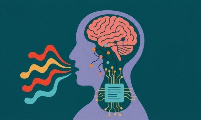
The cohort comprised 25 epilepsy patients temporarily implanted with stereo-EEG depth electrodes for clinical monitoring. During brief research sessions, participants read tongue twisters that stress the speech system and reduce guesswork about timing. The decoder targeted phonemes rather than whole words, aiming for a leaner path from neural activity to output text or synthesized voice.
“With it being a complex tongue twister task, your speech system is at a heightened alertness to minimize errors.”
That design choice mattered clinically. A phoneme-level model can learn transferable rules of articulation, then map those rules onto whatever viable language tissue a new patient still has. The group first built a shared recurrent layer that captured subject-independent features of speech planning and articulation, learned across thousands of channels placed through peri-sylvian language networks. They then froze that layer and adapted only lightweight parts of the network to each new person.
Why Transfer Learning Helps Patients Who Cannot Train Long
Training time is a scarce resource in hospital rooms. The study reports that a group-derived decoder consistently outperformed models trained on each individual alone, even when the new individual had limited electrode coverage or short recording sessions. In other words, prior patients loaned their neural priors. For someone with aphasia, that could mean achieving usable accuracy with minutes of calibration instead of days.
The mechanics are straightforward. Shared latent manifolds encode common articulatory structure across brains. A small subject-specific layer then learns how that person’s remaining network represents the same structure. Because the system decodes phoneme identity and position, it can operate before and during articulation, a property relevant to patients with very little overt speech.
The team also stress-tested what happens when key regions go missing. Occluding ventral sensorimotor cortex channels degraded performance the most in single-subject models, consistent with the central role of that strip in articulation. Group-pretrained models proved more resilient. For the clinic, resilience translates to a better chance of working around lesions, resections, or sparse coverage.
Picture the scene at the bedside. A gray cable bundle arcs from a compact amplifier. On a monitor, thin traces ripple as the patient prepares to say a hard sequence. The decoder, already steeped in patterns learned from others, needs only a few trials to align to this brain’s idiosyncrasies. The computer prints phonemes that quickly assemble into words. It is not magic, just better initialization.
From Lab Task To Bedside Tool For Aphasia
Patients with aphasia do not always have intact speech-motor cortex, fluent output, or the stamina for weeklong training. This strategy addresses all three constraints. It leverages multi-subject depth recordings to learn robust pre-articulatory and articulatory signatures of phoneme production, then fine-tunes minimally for the next user. The result is a decoder that starts closer to the finish line.
The study’s scope is technical but the implication is clinical. Hospital teams could maintain a growing library of neural features distilled from prior participants. When a new patient arrives, the system selects compatible priors, adapts in a short session, and begins assisting communication. That workflow would not replace therapy or long-form rehabilitation, but it could bridge a dangerous gap in which patients cannot express needs, symptoms, or consent.
Limits remain. The work used overt speech and a constrained stimulus set rather than spontaneous conversation. Inner speech was not analyzed, and real-world variability in prosody and volume needs attention. Still, the path forward is visible: broaden the language corpus, expand shared manifolds across centers, and validate rapid calibration in people with aphasia after stroke or brain injury.
“This means there is maximum engagement of the speech system and a lot of neural activity that we can tap into to decode what they are saying or trying to say.”
For now, the advance is a clean systems result with patient-facing potential. By pooling data across individuals, the field can make brain-to-text tools more accessible to those who need them most, not just those who can train the longest.
Nature Communications: 10.1038/s41467-025-63825-0
ScienceBlog.com has no paywalls, no sponsored content, and no agenda beyond getting the science right. Every story here is written to inform, not to impress an advertiser or push a point of view.
Good science journalism takes time — reading the papers, checking the claims, finding researchers who can put findings in context. We do that work because we think it matters.
If you find this site useful, consider supporting it with a donation. Even a few dollars a month helps keep the coverage independent and free for everyone.