A new technology is bringing real hope to spinal cord injury patients. Scientists at the University of Minnesota Twin Cities have combined 3D printing, stem cell biology, and organoid science to create scaffolds that restore nerve connections in rats with completely severed spinal cords. Published in Advanced Healthcare Materials, the study shows that 3D printed organoid scaffolds seeded with human stem cell–derived spinal neural progenitor cells (sNPCs) can integrate into damaged tissue, guide axon growth, and enable significant recovery of movement. While still in early stages, the research points toward a transformative treatment strategy for spinal cord injury, which currently has no cure.
Spinal Cord Injury and the Challenge of Repair
Spinal cord injuries affect more than 300,000 people in the United States, according to the National Spinal Cord Injury Statistical Center. Damage often leads to permanent paralysis because neurons die and nerve fibers cannot regrow across the lesion site. Existing therapies focus on preventing further harm and managing complications, but none fully restore lost function. Regenerative medicine approaches, particularly stem cell–based strategies, have long held promise but face hurdles in directing organized growth and ensuring integration with host neural circuits.
Printing a Scaffold for Regeneration
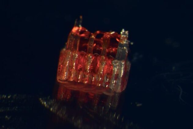
The Minnesota team developed a silicone scaffold just 2 millimeters long, containing microscale channels that mimic the architecture of the spinal cord. Using human induced pluripotent stem cells (iPSCs), they derived region-specific sNPCs and printed them into the channels with a supportive gel. In culture, these cells matured into spinal cord organoids—miniature, structured neural tissues—that exhibited diverse neuron types and functional activity for over a year.
“We use the 3D printed channels of the scaffold to direct the growth of the stem cells, which ensures the new nerve fibers grow in the desired way. This method creates a relay system that when placed in the spinal cord bypasses the damaged area.” —Guebum Han, former University of Minnesota postdoctoral researcher, now at Intel Corporation
Results in Rats
The researchers implanted two scaffolds into rats with fully transected spinal cords. Over 12 weeks:
- Neurons grew axons both rostrally and caudally, bridging the gap.
- New cells integrated with the host tissue and formed synapses.
- Rats showed marked improvements in locomotor scores compared to controls.
- Motor evoked potentials confirmed restored signal conduction through the injury site.
By the study’s end, most implanted cells had differentiated into neurons, while smaller populations became oligodendrocytes and astrocytes, replicating the cellular diversity of spinal tissue.
Clinical Potential and Next Steps
Senior author Ann Parr, professor of neurosurgery at the University of Minnesota, emphasized the long-term vision: “Regenerative medicine has brought about a new era in spinal cord injury research. Our laboratory is excited to explore the future potential of our ‘mini spinal cords’ for clinical translation.”
The team is now exploring biodegradable scaffolds and automated printing methods that can scale the approach for human-sized injuries. They also plan to incorporate dorsal sensory neurons to restore both motor and sensory functions.
Key Findings
- Sample: 18 female athymic rats with complete T8/9 transection; 5 received organoid scaffolds.
- Scaffold design: 3D printed silicone, 1.6 mm wide, 2 mm long, with three 200 µm channels.
- Cells: Human iPSC-derived region-specific spinal neural progenitor cells (sNPCs).
- Duration: In vitro maturation up to 365 days; in vivo study lasted 12 weeks post-transplantation.
- Effects: Significant locomotor recovery, restored motor evoked potentials, robust axonal growth and synapse formation.
- Location: University of Minnesota Twin Cities, with collaborators at Virginia Commonwealth University.
- Funding: NIH, Minnesota Spinal Cord Injury and TBI Research Grant Program, Spinal Cord Society.
- Safety: Human cells survived without immunosuppression in athymic rats; long-term human safety remains untested.
Takeaway
University of Minnesota researchers have created 3D printed spinal cord organoid scaffolds that restore nerve connections and mobility in rats with complete spinal cord transections. By combining printing technology with stem cell biology, the method offers a potential pathway toward human therapies for spinal cord injury.
Journal: Advanced Healthcare Materials
DOI: 10.1002/adhm.202404817
ScienceBlog.com has no paywalls, no sponsored content, and no agenda beyond getting the science right. Every story here is written to inform, not to impress an advertiser or push a point of view.
Good science journalism takes time — reading the papers, checking the claims, finding researchers who can put findings in context. We do that work because we think it matters.
If you find this site useful, consider supporting it with a donation. Even a few dollars a month helps keep the coverage independent and free for everyone.