A thin slice of human tissue sits in a laboratory dish, surrounded by nothing but a soft gel. But this isn’t just any tissue sample slowly dying on a lab bench.
For seven straight days, this piece of lymph node continues to function as if it were still inside a living person. It mounts immune responses, activates T cells, and even produces antibodies.
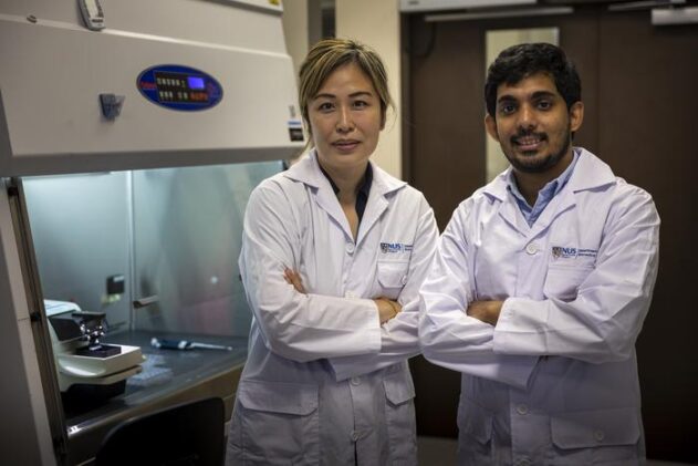
That’s the remarkable achievement described by researchers at the National University of Singapore and the National Cancer Centre Singapore. They’ve developed a method to keep human lymph node tissue alive and functional far longer than anyone thought possible.
The findings, published August 29 in Trends in Biotechnology, could transform how scientists study human immunity and develop new vaccines and cancer treatments.
“Lymph nodes are key to how our immune system detects and responds to disease, but they’ve been notoriously hard to study outside the body,” said Professor N Gopalakrishna Iyer, Senior Consultant at the National Cancer Centre Singapore and co-lead of the study.
Until now, lymph node tissue typically broke down within a day or two in laboratory conditions. This made detailed studies nearly impossible.
The Singapore team solved this by embedding thin slices of lymph node tissue from head and neck cancer patients into a specially designed hyaluronan-based hydrogel. This bioengineered scaffold mimics the body’s natural environment, supporting the tissue and dramatically reducing cell loss.
Beyond Simplified Models
The results speak to a fundamental problem in immunology research. Scientists have long relied on animal models or simplified cell cultures to understand human immunity. But these approaches often fail to capture the full complexity of how our immune system actually works.
Lymph nodes serve as command centers where immune cells gather to detect threats and coordinate responses. They’re those small, bean-shaped structures scattered throughout our bodies.
When the researchers exposed their lab-grown lymph node slices to cancer cells from the same patient, the tissue responded just as it would in the body. Immune cells became activated. Signaling molecules were released. The machinery of immunity kicked into gear.
Even more impressive: when exposed to a COVID-19 mRNA vaccine, the tissue samples produced specific immune responses and antibodies over several days.
One sample revealed something particularly intriguing. It showed an active immune response even before any vaccine exposure, likely reflecting the patient’s prior infection or vaccination history.
“This platform gives us a much more accurate picture of human immune responses, and how they can vary from person to person,” said Assistant Professor Eliza Fong from NUS, who co-led the study.
The Hydrogel Advantage
The secret lies in the hydrogel itself. In conventional setups, roughly 60% of cells flee from lymph node tissue within the first two days. This leaves researchers with degraded samples that bear little resemblance to functional immune tissue.
But the hydrogel-embedded samples maintained over 90% of their original size and cellular composition throughout the week-long study period.
The researchers found that less than 15% of cells migrated out of their hydrogel-supported tissue. Compare that to about 80% cell loss in traditional floating cultures. The hydrogel appears to act as both a physical barrier and a supportive scaffold, preventing the normal cellular exodus that dooms most tissue culture attempts.
While the current system supports tissue function for about a week, the team is working to extend that timeframe. They’re also adding features like lymph flow.
The real excitement lies in what this technology could unlock for personalized medicine. Rather than relying on generalized models, doctors might one day test how a patient’s own immune tissue responds to specific treatments before beginning therapy.
The researchers envision using this system for screening cancer vaccines, infectious disease treatments, and immunotherapies. It could provide a faster, more human-relevant alternative to animal testing while offering insights into the profound variability in how different people’s immune systems respond to the same threats.
But perhaps the most significant implication is what this means for our understanding of human immunity itself. For the first time, scientists can watch human immune responses unfold in real time, in tissue that maintains its natural architecture and cellular relationships.
That’s a window into human biology that researchers have never had before.
ScienceBlog.com has no paywalls, no sponsored content, and no agenda beyond getting the science right. Every story here is written to inform, not to impress an advertiser or push a point of view.
Good science journalism takes time — reading the papers, checking the claims, finding researchers who can put findings in context. We do that work because we think it matters.
If you find this site useful, consider supporting it with a donation. Even a few dollars a month helps keep the coverage independent and free for everyone.