For the first time, researchers have directly observed the tiny protein clusters believed to kickstart Parkinson’s disease in human brain tissue, a development one scientist likens to seeing stars during the day.
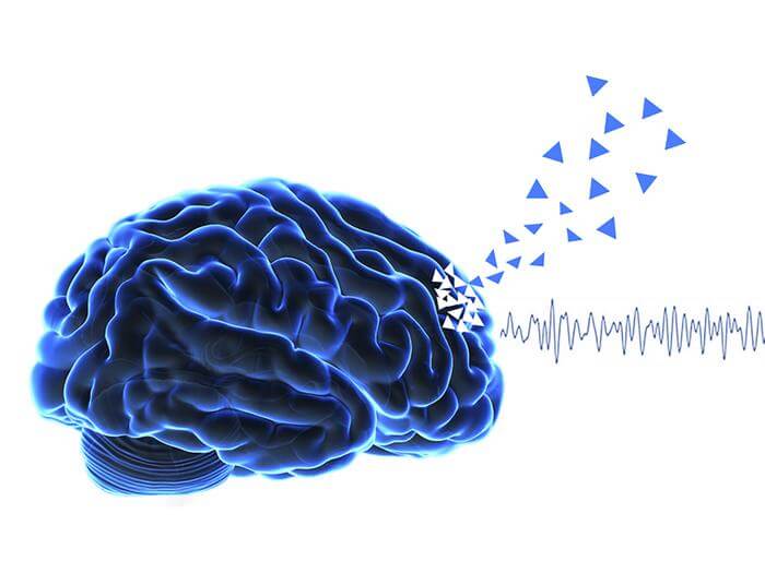
The clusters, called alpha-synuclein oligomers, measure just a few nanometers across and have eluded detection in human brains until now, despite being long suspected as the initial trigger for the world’s fastest-growing neurological disease. A team from the University of Cambridge, UCL, the Francis Crick Institute, and Polytechnique Montreal developed an imaging technique that finally makes these molecular needles visible in the biological haystack.
Their findings, published in Nature Biomedical Engineering, analyzed roughly 1.2 million protein aggregates from post-mortem brain tissue. The work could reshape how doctors diagnose and eventually treat a condition that currently affects 166,000 people in the UK alone, with global cases expected to double to 25 million by 2050.
Seeing What Was Always There
The new technique, ASA-PD (Advanced Sensing of Aggregates for Parkinson’s Disease), uses ultra-sensitive fluorescence microscopy to detect oligomers that standard methods simply cannot see. The challenge has always been one of scale. Doctors have recognized Parkinson’s for over a century through large protein deposits called Lewy bodies, but these structures tell you where the disease has been, not where it is right now.
“This is the first time we’ve been able to look at oligomers directly in human brain tissue at this scale: it’s like being able to see stars in broad daylight. It opens new doors in Parkinson’s research.”
Dr. Rebecca Andrews, co-first author who conducted the work as a postdoctoral researcher, captures the magnitude of what the team accomplished. The oligomers produce such weak signals that detecting them required maximizing the fluorescent signal while dramatically reducing background noise from brain tissue.
When the team compared brain tissue from Parkinson’s patients with healthy individuals of similar age, they found something unexpected. Oligomers exist in both groups. The critical difference was not presence or absence, but character. In diseased brains, the oligomers were larger, brighter, and far more numerous.
A Disease-Specific Signature
The team discovered a subset of oligomers appearing only in Parkinson’s patients. These could represent the earliest visible markers of disease, potentially years before symptoms emerge. The finding challenges assumptions about what triggers neurodegeneration and when.
Professor Steven Lee from Cambridge’s Yusuf Hamied Department of Chemistry, who co-led the research, notes the technique does not just provide a snapshot. Similar technologies could extend to other neurodegenerative diseases like Alzheimer’s and Huntington’s, conditions that share protein aggregation as a common thread but lack tools to observe it at this scale.
“Oligomers have been the needle in the haystack, but now that we know where those needles are, it could help us target specific cell types in certain regions of the brain.”
The researchers examined tissue from the anterior cingulate cortex, generating over 13,000 high-resolution images covering more than 41 square millimeters. They found that oligomers in Parkinson’s brains were not randomly distributed but clustered around specific cell types, including neurons, astrocytes, and microglia. Curiously, they were dispersed away from oligodendrocyte nuclei, suggesting some cells may be protected or simply uninvolved in the aggregation process.
The spatial analysis revealed another layer of complexity. While microscale Lewy bodies predominate in neurons, these smaller precursor oligomers appeared in and around multiple cell types. Whether they transition from physiological assemblies to pathological ones within neurons, or travel between cells, remains unclear. The team detected approximately 16 times more small aggregates than large ones in diseased tissue, underscoring that the oligomers represent the dominant form of protein accumulation.
Professor Sonia Gandhi from The Francis Crick Institute, who co-led the work, emphasizes that understanding human disease requires studying human brains directly, despite their complexity. Breaking through the technological barrier may now allow researchers to understand why, where, and how protein clusters form and how this changes the brain environment to cause disease.
The implications extend beyond basic science. No drugs currently slow or stop Parkinson’s itself. Treatments only manage symptoms like tremor and stiffness. If researchers can observe the disease at its earliest molecular stages, they may identify intervention points long before irreversible damage occurs. The dataset, metadata, and analysis tools have been made publicly available online, inviting the broader scientific community to explore the findings.
The research was supported by Aligning Science Across Parkinson’s (ASAP), the Michael J. Fox Foundation, and the Medical Research Council. The authors acknowledge the patients, families, and caregivers who donated tissue to brain banks, making this work possible.
Nature Biomedical Engineering: 10.1038/s41551-025-01496-4
ScienceBlog.com has no paywalls, no sponsored content, and no agenda beyond getting the science right. Every story here is written to inform, not to impress an advertiser or push a point of view.
Good science journalism takes time — reading the papers, checking the claims, finding researchers who can put findings in context. We do that work because we think it matters.
If you find this site useful, consider supporting it with a donation. Even a few dollars a month helps keep the coverage independent and free for everyone.