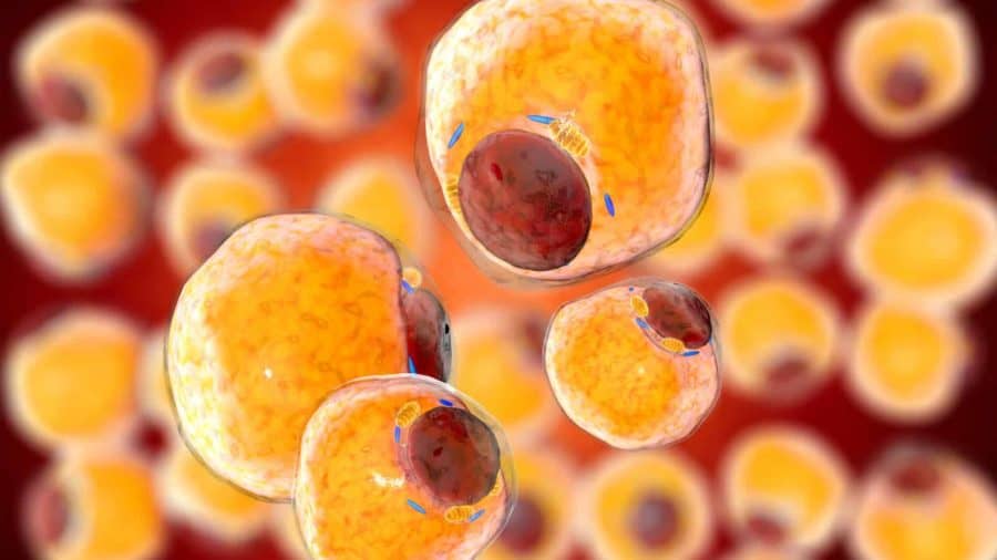
Fat isn’t glamorous. We vacuum it out during liposuction, toss it as medical waste, think of it as the body’s storage locker. But researchers in Shanghai saw something else: a living factory that could be repurposed without the usual genetic rewiring. They took ordinary adult adipose tissue and coaxed it into becoming bone marrow, insulin-secreting clusters, even neural tissue, all without isolating stem cells or touching a single gene.
The study, published in Engineering by a team at Shanghai Jiao Tong University School of Medicine, describes a surprisingly direct process. Human fat was mechanically processed into microfat, then cultured in suspension where it spontaneously condensed into what the researchers call “reaggregated microfat” or RMF pellets. Over weeks, these pellets transformed from a loose, yellowish slurry into smooth, firm spheres—like tiny pearls of reorganized tissue. The key wasn’t breaking the fat down to individual cells but letting it keep its internal structure, its “neighborhood,” which helped cells shift into new identities.
Diabetic Mice, Reversed
The most dramatic test came with diabetes. The team guided RMF pellets down an endodermal path using a four-stage protocol, eventually creating islet organoids that produce insulin. When these organoids were transplanted into diabetic mice, they didn’t just survive—they integrated. Within two weeks, the mini-organs had tapped into the animals’ blood supply and begun secreting insulin in response to glucose. Blood sugar levels normalized and stayed that way for the duration of the experiment.
It’s a result that underscores the functional potential here. These weren’t decorative tissue models; they were working organs performing the exact job pancreatic islets do in a healthy body. The researchers observed the RMF pellets condensing and remodeling their extracellular matrix in ways typically associated with regenerative niches—environments where cells naturally repair and rebuild.
“These findings therefore highlight the potential of human adipose tissue as a safe, scalable, and clinically viable source for organoid-based regenerative therapies,” Qingfeng Li explains.
The same starting material proved equally versatile in other directions. When pushed toward mesoderm, RMF pellets underwent endochondral ossification in immunodeficient mice, forming humanized bone marrow organoids complete with the specialized niches that support blood cell production. After receiving human stem cells, these tiny bone shells sustained long-term hematopoiesis—a functional marrow environment grown from fat. For ectoderm, the pellets formed neurospheres and differentiated into cells expressing neuronal and glial markers, demonstrating neural-like tissue entirely in vitro.
Bypassing the Stem Cell Bottleneck
Traditional organoid methods are slow and expensive. They rely on pluripotent stem cells that must be isolated, expanded, and meticulously guided through differentiation. That process is labor-intensive, hard to scale, and carries risks like tumor formation when reprogrammed cells misbehave. The RMF approach sidesteps all of that by preserving the tissue’s natural architecture. Instead of dismantling fat into single cells, the method allows it to reorganize itself using the signaling already embedded in its structure.
Because adult fat is abundant and easy to obtain—often discarded during routine procedures—this technique could scale far more readily than stem cell protocols. It also opens a personalized medicine angle: a patient’s own fat could theoretically be used to grow cells tailored specifically to their immune system, minimizing rejection risk. The researchers showed that RMF pellets increased cell proliferation and reorganized their matrix in ways that mimic regenerative environments, suggesting the tissue retains a surprising degree of plasticity even in adulthood.
The study doesn’t claim this is ready for the clinic. More testing is needed, particularly around long-term safety and the consistency of differentiation across different donors. But the principle is clear: fat tissue, when handled correctly, behaves less like a fixed cell type and more like flexible raw material. It’s a conceptual shift that could make organoid production faster, cheaper, and more accessible—turning what was once waste into a cornerstone of regenerative medicine.
Engineering: 10.1016/j.eng.2025.06.031
Inside the Science: How Fat Becomes a Lab-Grown Organ
The transition from a patient’s “love handles” to a functioning medical organoid involves a sophisticated mechanical and biological sequence. Unlike traditional methods that break tissue down into individual cells, this technique preserves the “neighborhood” of the cells, known as the extracellular matrix (ECM).
The RMF Process: From Fat to Pellets
The journey begins with lipoaspirate—the fat removed during liposuction. Instead of discarding it, scientists process it into Reaggregated Microfat (RMF). The tissue is rinsed and mechanically refined to create a concentrated slurry of cells and their supporting scaffold. These are then placed into a specialized suspension culture where they naturally self-assemble into 4-millimeter “RMF pellets.” These pellets act as the blank canvas for any organ the scientists wish to grow.
Three Paths of Transformation
Once the pellets are formed, the team uses specific chemical signals to push them down different developmental paths, representing the three germ layers of an embryo:
- The Endoderm Path (Islets): To treat diabetes, the pellets are exposed to growth factors that mimic pancreatic development. Over four stages, the fat cells transform into insulin-secreting beta cells. These clusters, or organoids, are then able to monitor glucose levels and release insulin just like a healthy pancreas.
- The Mesoderm Path (Bone Marrow): Pellets are treated with “chondrogenic” (cartilage-forming) media. Once implanted in the body, these pellets undergo a process called endochondral ossification. They harden into bone and develop a hollow interior that becomes home to a humanized bone marrow environment.
- The Ectoderm Path (Neural Tissue): By using a different chemical cocktail, the researchers can “reprogram” the pellets to express neural markers. This results in neurospheres that contain the building blocks of the central nervous system, including neurons and support cells (glia).
Why Adipose Tissue?
Adipose tissue is uniquely suited for this work because it is rich in Mesenchymal Stem Cells (MSCs) and pericytes—cells that are naturally designed to repair and maintain blood vessels. By keeping these cells in their native environment during the RMF process, scientists ensure the resulting organoids are “pre-vascularized,” meaning they are ready to connect to a patient’s blood supply almost immediately after transplantation.
ScienceBlog.com has no paywalls, no sponsored content, and no agenda beyond getting the science right. Every story here is written to inform, not to impress an advertiser or push a point of view.
Good science journalism takes time — reading the papers, checking the claims, finding researchers who can put findings in context. We do that work because we think it matters.
If you find this site useful, consider supporting it with a donation. Even a few dollars a month helps keep the coverage independent and free for everyone.