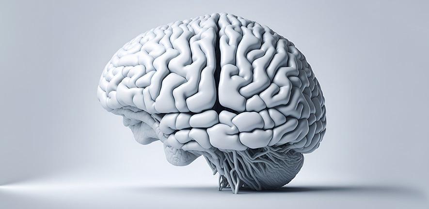
Inside every cell, tiny molecular machines are running more or less continuously, churning out a molecule called adenosine triphosphate. ATP is the basic unit of biological energy, the chemical that powers almost everything from a muscle contraction to a thought, and the mitochondria that make it are perpetually adjusting their output to match demand. In a healthy cell, the system is tuned, responsive, capable of ramping up when the body needs it. In the cells of young adults with major depressive disorder, something rather different is happening.
A new study published in Translational Psychiatry suggests that in early-stage depression, these cellular energy systems are already running close to their ceiling. And that near-maximal effort at rest, while it keeps baseline function going, may be precisely what makes fatigue so crippling.
The finding, from researchers at the University of Queensland and the University of Minnesota, is the first to measure ATP dynamics simultaneously in the living brain and in blood cells of young people with depression. That pairing matters more than it might seem. Previous research has occasionally flagged energy metabolism as abnormal in depression, but it has generally looked at the brain alone, and it has measured ATP concentration rather than rate of production. The difference between those two things is a bit like the difference between checking how much petrol is in your tank versus checking how hard the engine is working. You can have a full tank and a struggling engine, and that, it seems, is something like what’s happening here.
The brain imaging relied on a technique developed at the University of Minnesota called phosphorus-31 magnetic resonance spectroscopy with magnetization transfer, run at 7 Tesla, which is roughly four times the field strength of a standard hospital scanner. What that buys you is the ability to measure not just the amount of ATP in the visual cortex but the actual rate at which mitochondria are synthesising it. The team scanned nine young adults with major depressive disorder and nine healthy controls, all between 18 and 24 years old, and on the same day drew blood to measure ATP metabolism in immune cells from each participant.
The results surprised them. The expectation going in, reasonably enough, was that depressed participants might show sluggish energy production. What they actually found was the opposite. “This was surprising,” said Dr. Roger Varela of the Queensland Brain Institute, “because you might expect energy production in cells would be lower for people with depression.” The brain cells of the depressed participants were producing ATP faster than those of the healthy controls, not more slowly. Their blood cells held higher resting concentrations of ATP too.
On its face, that looks like a discrepancy with the lived experience of depression, where exhaustion is one of the most common and treatment-resistant complaints. But the second part of the measurement is what ties the story together. When the research team chemically stressed the blood cells, forcing the mitochondria to work harder than they would at rest, the depressed group’s cells fell behind. They couldn’t ramp up oxygen consumption to match, which meant their capacity reserve, the headroom available for higher energy demands, was substantially reduced. As Varela put it, “it suggests that in the early stages of depression, the mitochondria in the brain and body have a reduced capacity to cope with higher energy demand, which may contribute to low mood, reduced motivation and slower cognitive function.”
The picture that emerges, then, is a compensatory one. The mitochondria in early-stage depression appear to be compensating for some underlying deficit by running at elevated output when the body is at rest. That compensation keeps baseline function roughly intact, and it’s why the resting ATP concentration in the brain looks normal, actually identical between the two groups in this study. But it comes at a cost: it leaves the system with little room to respond when additional demand arrives. Fatigue, under this model, isn’t a deficiency so much as a ceiling. The cells are doing what they can; they’ve just already done most of it.
That the same signal showed up in both the brain and the blood is what makes the study stand out. “This shows multiple changes occur in the body, including in the brain and the blood, and that depression impacts energy at a cellular level,” said Varela. And the two measures correlated with each other, meaning participants with higher ATP production rates in their visual cortex tended also to have higher ATP concentrations in their blood. Whether blood ATP could eventually serve as a proxy measure for brain energetics, which would be considerably cheaper and easier to obtain than a 7 Tesla brain scan, is one of the more intriguing questions the work opens up.
The study is explicitly preliminary. The sample is small (9 participants per group for brain imaging; 11 and 13 for blood), it wasn’t balanced for sex, and many of the depressed participants were taking psychotropic medications, which is realistic but introduces noise. After correcting for age and sex, the brain imaging findings dropped from statistically significant to trend-level, so larger, better-controlled replication will be needed before anyone calls this settled. Associate Professor Susannah Tye, also from the Queensland Brain Institute, was careful not to overstate it: “Fatigue is a common and hard-to-treat symptom of MDD, and it can take years for people to find the right treatment for the illness. There has been limited progress in developing new treatments because of a lack of research and we hope this important breakthrough could potentially lead to early intervention and more targeted treatments.”
One detail the researchers note in passing is worth dwelling on. The same elevated-ATP-production-rate signature in the visual cortex has been found before, in patients with early-stage Parkinson’s disease, using the same imaging method. That could be coincidence, or it could be pointing to something more general: a common compensatory response in the early phase of neurological disease, before the system starts to fail. The speculative extension is that this failure, if it comes, might eventually look like the declining ATP concentrations that older studies found in chronically depressed adults; the early upswing might be the system still fighting. Depression in early life also increases the risk for dementia later on. Whether the energy dynamics seen here are part of that pathway is not something this study can address, but it is the kind of question the methodology is now, for the first time, sharp enough to ask.
“This suggests that depression symptoms may be rooted in fundamental changes in the way brain and blood cells use energy,” Tye said. If the compensatory mechanism can be detected early, perhaps through a blood test calibrated to the ATP biosignature described here, the goal shifts from treating established disease to catching the engine before it runs out of headroom.
DOI / Source: https://doi.org/10.1038/s41398-026-03904-y
Frequently Asked Questions
The paradox dissolves when you look at what the cells do under stress rather than at rest. In early depression, mitochondria appear to compensate for an underlying deficit by running at elevated output during normal activity, keeping baseline function intact but leaving little reserve for higher demands. When those demands arrive, the system can’t respond, and that ceiling, rather than a simple shortage, may be what registers as fatigue.
Possibly, though the research is at a very early stage. The study found that ATP levels in blood cells correlated with both depression severity and fatigue scores, and also correlated with the brain’s own ATP production rate. If that relationship holds up in larger studies, a blood-based ATP measurement might eventually serve as a cheaper and less invasive proxy for what’s happening in the brain itself.
Earlier brain imaging studies of depression could measure how much ATP was present in brain tissue, but not how quickly it was being made. The 7 Tesla phosphorus-31 technique used in this study captures the rate of ATP synthesis, which turns out to be the more informative number. Normal resting concentrations can coexist with a hidden capacity problem, and only the rate measurement reveals that.
Not exactly, though the findings push in that direction. What the study suggests is that the symptoms of depression, particularly fatigue, reduced motivation, and cognitive slowing, may have a cellular energy basis that sits beneath, or alongside, the neurochemical explanations most treatments currently target. Whether that metabolic layer is a cause or a consequence of depression remains an open question.
The researchers speculate, carefully, that the elevated energy production seen in these young adults with early-stage depression may represent a temporary compensation. In older studies of people with long-standing depression, ATP levels in blood cells were lower than normal rather than higher. If that picture holds, the early upswing might eventually give way to a collapse, though longitudinal studies tracking the same patients over years would be needed to know whether that trajectory is real.
ScienceBlog.com has no paywalls, no sponsored content, and no agenda beyond getting the science right. Every story here is written to inform, not to impress an advertiser or push a point of view.
Good science journalism takes time — reading the papers, checking the claims, finding researchers who can put findings in context. We do that work because we think it matters.
If you find this site useful, consider supporting it with a donation. Even a few dollars a month helps keep the coverage independent and free for everyone.
Key Takeaways
- A recent study finds that young adults with major depressive disorder show heightened ATP production in brain and blood cells.
- Despite increased ATP output, these individuals struggle with fatigue due to a reduced capacity to cope with energy demands.
- The study measured ATP rates using a 7 Tesla scanner, revealing important insights into energy dynamics in depression.
- The findings suggest a cellular basis for depressive symptoms, including fatigue and cognitive slowing, potentially opening avenues for early intervention.
- Further research is needed to confirm these results and explore blood tests as a proxy for brain energy dynamics.