Inside every cell exists a bustling world of molecular machinery, where proteins and RNA molecules zip around carrying out the instructions encoded in our DNA. But scientists have discovered that some of this traffic gets stuck in peculiar nanoscale traps, and understanding these tiny detention centers might help explain what goes wrong in devastating diseases like ALS.
Researchers at the University of Michigan have developed a method to peer inside droplet-like structures called biomolecular condensates, revealing for the first time that molecules do not move freely within them. Instead, proteins and RNA get confined in distinct zones the team calls “nanodomains,” infinitesimal regions that slow molecular movement to a crawl.
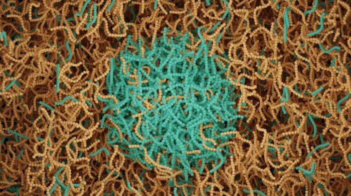
The finding challenges the prevailing view of condensates as uniform liquid droplets. They are more like cities with distinct neighborhoods, each with its own traffic patterns and chemistry.
“There is a lot of hope that by manipulating these condensates, we can use them for medical purposes, such as slowing neurodegenerative disease, making them a repository for drugs that can be released slowly over time, or sequestering unwanted proteins such as those that are cancer- or virus-related by inducing them to form condensates,” said Nils Walter, director of the Center for RNA Biomedicine at U-M and senior author of the study.
Wrestling Droplets Into Focus
The research team focused on a protein called fused in sarcoma, or FUS, which forms condensates under cellular stress. When mutated, FUS accumulates in clumps linked to ALS and frontotemporal dementia. But watching these processes unfold has been maddeningly difficult.
The main problem? The droplets will not sit still. Place one on a microscope slide and it rolls around like a marble, making precise imaging nearly impossible. Add too many molecular anchors to hold it in place and the spherical droplet flattens into what Walter calls “a pancake.”
The team found the sweet spot: just enough anchors to immobilize the condensates while preserving their three-dimensional structure. They then tagged individual FUS proteins and RNA molecules with fluorescent dyes, creating a tracking system that could follow single molecules as they moved through the droplets.
What emerged from thousands of molecular trajectories was unexpected. Rather than distributing evenly, the tracked molecules clustered in specific regions, leaving other areas relatively empty. The pattern revealed nanodomains: zones roughly 300 nanometers across where diffusion slows dramatically.
Different Molecules, Different Neighborhoods
The researchers discovered that FUS proteins and RNA molecules occupy separate nanodomains that actively exclude each other. About 67% of FUS proteins and 83% of RNA molecules showed confined movement, compared to just 40% of RNA floating freely outside the condensates.
Size and shape matter tremendously in this nanoscale world. Long messenger RNA strands, about 1,500 nucleotides, got trapped readily in nanodomains. Shorter microRNA molecules moved more freely. Tiny fluorescent dye molecules barely got confined at all.
The team tested spherical beads with the same size and electric charge as RNA. The beads got stuck even more than RNA, suggesting that RNA’s flexible, thread-like structure lets it squeeze through the condensate’s molecular mesh.
“If you make a condensate and put it on a microscope slide, it can roll over the surface or wiggle back and forth. If that happens, then the particle tracking gets messed up. So you have to immobilize the condensates on the surface, but you have to do that in a very judicial way.”
Electric charge played a surprising role too. Positively charged and negatively charged nanoparticles moved similarly, but neutral particles got immobilized more often. The findings suggest nanodomains act less like rigid cages and more like tangled nets that selectively trap certain molecular shapes and sizes.
This selective trapping has consequences. An RNA molecule might dart freely through a condensate until it encounters a nanodomain, where it gets temporarily detained. One tracked RNA molecule spent nearly two seconds stuck in two different nanodomains before finally reaching the droplet edge and escaping. That extended residence time could influence whether the RNA gets processed, degraded, or protected.
As the condensates aged over 24 hours, the nanodomains began migrating toward the droplet surface. There, the researchers observed something striking: protein fibrils, the toxic aggregates associated with ALS, sprouted from the condensate surface. The nanodomains appeared to seed this transformation from liquid droplet to solid aggregate.
The team then tested two FDA-approved ALS drugs, edaravone and riluzole, to see how they affected nanodomain behavior. Both drugs accelerated the movement of nanodomains to the condensate surface and sped up fibril formation. The effect was particularly pronounced with riluzole.
At first glance, promoting fibril formation seems counterintuitive for an ALS treatment. But some researchers theorize that larger, stable fibrils might actually protect neurons by soaking up smaller, more toxic protein aggregates. If correct, the drugs may work partly by hurrying this defensive response along.
“But what our findings mean overall is that, for the first time, we see these nanodomains as potential seeds to these fibers. Maybe the drugs we used, edaravone and especially riluzole, have another effect beyond those known, by helping the condensates to fibralize faster and protect the neuron.”
The discovery opens new questions about how cells normally regulate these condensates and what triggers their transformation into disease-causing aggregates. The nanodomains appear within minutes of condensate formation, suggesting they are fundamental features rather than signs of aging or damage.
Understanding condensate architecture could inform strategies for treating not just ALS but other diseases involving protein aggregation, from Alzheimer’s to certain cancers. It might also aid efforts to engineer artificial condensates for drug delivery, where controlling how long medications remain trapped could determine treatment effectiveness.
The field of biomolecular condensates has exploded in recent years as researchers recognize how many cellular processes depend on these membraneless compartments. But most studies have treated them as homogeneous liquids. The new findings reveal a far more complex internal geography, one that actively shapes the behavior and fate of molecules passing through.
For now, the Michigan team’s technique works only with purified proteins in controlled laboratory conditions. Cells present additional challenges: more crowded environments, multiple types of condensates, and constant reorganization. But the basic principles likely apply across different proteins and cellular contexts.
The research, supported by the National Institutes of Health, National Science Foundation, and Chan Zuckerberg Initiative, appears in Nature Nanotechnology. It offers a reminder that even in structures as small as cellular droplets, invisible to any microscope just decades ago, there exist layers of organization that profoundly affect human health and disease.
Nature Nanotechnology: 10.1038/s41565-025-02077-x
ScienceBlog.com has no paywalls, no sponsored content, and no agenda beyond getting the science right. Every story here is written to inform, not to impress an advertiser or push a point of view.
Good science journalism takes time — reading the papers, checking the claims, finding researchers who can put findings in context. We do that work because we think it matters.
If you find this site useful, consider supporting it with a donation. Even a few dollars a month helps keep the coverage independent and free for everyone.