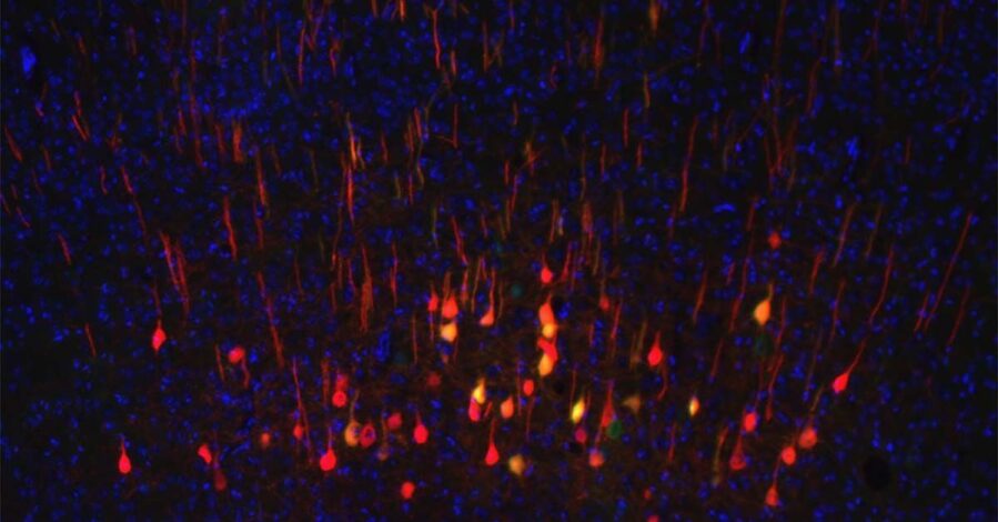
In some parts of the aging brain, the opposite of what you might expect happens. Even as brain tissue shrinks, the remaining cells start talking to each other more, not less. By studying brain scans from nearly 28,000 adults, researchers found that visual areas often ramp up their internal communication to make up for physical loss. This extra coordination appears to help protect memory and problem-solving skills, even as brain volume declines.
The finding pushes back against the idea that brain aging is a steady slide downhill. Instead, scientists at Shanxi University and Georgia Tech saw two different patterns unfolding at once. Some brain networks weaken across the board, losing both structure and signal. Others seem to work harder, strengthening connections to keep performance steady despite physical wear. These opposing trends only became clear when the team examined brain structure and brain activity together, rather than looking at either one on its own.
Using data from the UK Biobank, the team combined gray matter volume measurements with functional connectivity scans that track how strongly different brain regions communicate at rest. Models trained on both data types predicted a person’s age far more accurately than either measure alone, revealing that the relationship between structure and function carries information that neither captures independently.
Where Decline Doubles Down
In many brain regions, the aging process follows what researchers call a synergistic pattern. The cerebellum, frontal pole, and areas involved in attention and high-level thinking showed coordinated declines in both tissue volume and communication strength. When these regions take a double hit, the consequences show up in behavior. Weakening connections between the cerebellum and paracingulate gyrus, for instance, tracked closely with slower reaction times in study participants.
This synchronized deterioration makes intuitive sense: less tissue means fewer neurons to maintain strong signals. But it is not the whole story. The contradictory pattern, where function increases as structure decreases, emerged most clearly in the occipital cortex and other visual processing areas. As these regions lost gray matter, their internal connectivity strengthened, and people with more pronounced compensation in these areas scored better on tests of fluid intelligence and numeric memory.
“Aging primarily induces synergistic changes, with both functional connectivity and gray matter volume decreased, but also contradictory changes that act as a compensatory mechanism as one ages,” Yuhui Du explains.
The dataset’s size allowed the researchers to detect lateralization effects that smaller studies miss. The right thalamus, a sensory relay station, showed increasing volume with age, while the left side tended to shrink. These left-right asymmetries suggest that even adjacent brain structures can age along different trajectories, complicating efforts to develop one-size-fits-all interventions.
Resilience Has Limits
The compensation mechanism offers a more optimistic take on cognitive aging than the decline-only model, but it is not infinite. Visual regions can turn up their communication to offset moderate tissue loss, but there is presumably a threshold beyond which no amount of increased connectivity can maintain function. The study does not identify where that breaking point lies, though it hints that early detection might focus less on tissue volume alone and more on whether compensatory patterns remain intact.
For clinicians, the findings suggest that brain health assessments could become more nuanced. Instead of flagging shrinkage as an automatic warning sign, doctors might look for breakdowns in the relationship between structure and function. A brain that is shrinking but maintaining strong connectivity might be in better shape than one where both are declining together, even if the total tissue loss appears similar on a standard MRI.
The research frames aging not as a passive slide toward dysfunction but as an active process where the brain reorganizes itself in response to physical constraints. That reframing could reshape how we think about interventions, shifting focus from preventing tissue loss (which may be inevitable) to supporting the brain’s compensatory mechanisms that keep cognition stable despite that loss.
Research: 10.34133/research.0887
ScienceBlog.com has no paywalls, no sponsored content, and no agenda beyond getting the science right. Every story here is written to inform, not to impress an advertiser or push a point of view.
Good science journalism takes time — reading the papers, checking the claims, finding researchers who can put findings in context. We do that work because we think it matters.
If you find this site useful, consider supporting it with a donation. Even a few dollars a month helps keep the coverage independent and free for everyone.