Start with a network map and you can see something odd. A cascade of molecular connections threads outward from a handful of membrane proteins, touching enzymes that process purines, converging on G-protein subunits that normally sit quiet until a chemical signal triggers them.
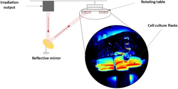
The network isn’t pathological, exactly. It looks, if anything, like a cell running a controlled drill. Something arrived from outside, the membrane registered it, and a coordinated biochemical response rippled inward without a single cell dying. The “something” was terahertz radiation: electromagnetic waves at 2.3 terahertz, produced by a massive free-electron laser in Novosibirsk, aimed at human melanoma cells for 45 minutes.
The results, published in Gene Expression by a Siberian team at the Institute of Cytology and Genetics, are interesting less for what terahertz radiation destroys than for what it quietly rearranges.
Terahertz radiation sits between infrared and microwave in the electromagnetic spectrum, roughly 0.1 to 10 terahertz. It passes through many biological materials without ionizing them, unlike X-rays, which makes it potentially useful for imaging and therapy. Interest has grown because terahertz frequencies correspond to natural vibrational modes of biological molecules, meaning the radiation interacts with cells in a structurally specific rather than simply thermal way.
The purine salvage pathway recycles existing nucleotide fragments into new DNA and RNA building blocks, using far less ATP and fewer amino acids than building nucleotides from scratch. Cells favor salvage when energy or raw materials are constrained. In this study, the shift appears to be an adaptive response to terahertz-induced membrane changes rather than a sign of cellular stress, since cell viability remained high throughout.
Lipid rafts are cholesterol-rich regions within cell membranes that organize signaling proteins into functional clusters. They are thought to be sensitive to terahertz radiation because the collective molecular motions of phospholipid bilayers and their surrounding water layers occur at precisely the terahertz frequency range, giving the radiation a physical coupling to membrane structure. Disrupting or rearranging rafts would scatter or concentrate the signaling proteins they host, potentially triggering downstream cellular responses.
Melanoma is considered a promising candidate for terahertz-based approaches partly because skin tumors are directly accessible, avoiding the tissue-penetration challenges of reaching deeper cancers. Some earlier research suggests terahertz frequencies can influence DNA methylation, which is abnormal in many melanomas. But this study is preclinical and mechanistically theoretical: a single cell line, a single frequency, conclusions based on network modeling rather than direct experimental intervention. Clinical applications remain speculative.
Terahertz radiation occupies an awkward gap in the electromagnetic spectrum, between infrared and microwave, and for a long time that awkwardness extended to the biology. The waves interact with tissue, earlier studies had shown membrane effects, gene expression changes, various cellular responses, but the underlying mechanism stayed murky. What makes interpretation difficult is that terahertz radiation can work through two distinct routes. At high power it heats tissue; at the lower powers used in Novosibirsk, it doesn’t heat anything measurable at all. Whatever is happening must therefore be non-thermal, meaning something structurally specific is going on at the molecular level rather than the biological equivalent of warming cells in a bath.
The membrane is probably where it starts. Phospholipid bilayers and their hydration shells support collective molecular motions in precisely the terahertz frequency range, giving the radiation a direct physical handle on membrane structure. Within membranes sit lipid rafts: dynamic cholesterol-rich microdomains that concentrate signaling proteins and act as organizing platforms for cellular communication. The Novosibirsk team’s hypothesis, built on their earlier work, was that terahertz radiation perturbs these rafts, and that this perturbation sets off the metabolic changes they were detecting.
To test it properly they needed two things: a chemical readout of what the cells were actually doing, and a way to connect those chemical changes back to the membrane. The first came from a high-resolution mass spectrometry metabolomics screen that identified 40 significantly altered metabolites in the irradiated cells. The second came from gene network reconstruction using a bioinformatics platform called ANDSystem, which mines the scientific literature and protein databases to rebuild regulatory connections between proteins. Together they traced a path from the membrane outward to the metabolites and back.
The metabolomics revealed a recognizable pattern. The biggest shift was in purine metabolism (a p-value of roughly 10 to the power of minus 11, about as unambiguous as biological data gets). Adenine levels fell. Hypoxanthine, inosine triphosphate, and deoxyinosine rose. To a cell biologist, that combination has a clear signature: it’s a shift from de novo purine synthesis toward the purine salvage pathway. De novo synthesis is expensive, requiring considerable ATP and amino acids. Salvage is thriftier, recycling existing nucleotide fragments rather than rebuilding them from scratch. The cell appears to have switched to a more economical energy mode.
The gene network analysis offers a possible mechanism. The enzyme adenosine deaminase, which converts adenosine to inosine in the first step of the salvage cascade, forms direct physical contacts with five membrane receptors, including DPP4 (a protein familiar to diabetes researchers, but also active on tumor cell surfaces). DPP4 is known to anchor and activate adenosine deaminase precisely in lipid rafts. If terahertz-induced raft rearrangement increases DPP4 activity, the researchers reason, that would accelerate the salvage cycle and produce the metabolite pattern observed. It’s a plausible story, and notably an adaptive one: the cell isn’t breaking down, it’s adjusting its energy bookkeeping in response to an unfamiliar stimulus.
Three G-protein subunits, GNAI1, GNAQ, and GNAS2, showed up in the network as extraordinarily well-connected nodes, each receiving regulatory inputs from 11 to 29 different plasma membrane receptors. These proteins act, in the team’s framing, as a kind of molecular gateway: the more signals converge on the G-alpha nodes, the stronger the push toward salvage activity and away from the costlier de novo route. The epidermal growth factor receptor (EGFR) appeared too, connected to enzymes governing nucleotide reutilization. Notably, the EGFR clustering apparently didn’t require any growth factor ligand; earlier work has shown that physical perturbations of lipid rafts can activate EGFR without any chemical signal at all.
Two other pathways shifted alongside purines: pantothenate and CoA biosynthesis, important for mitochondrial energy transfer, and the pentose phosphate pathway, which generates NADPH for antioxidant defense. Neither was as dramatic as the purine shift, but together they suggest a coordinated metabolic adjustment aimed at maintaining redox balance under electromagnetic stimulation.
Throughout all of this, the cells survived. Seventy-two hours after irradiation, viability was 96.6 percent, essentially indistinguishable from 97.6 percent in controls. That’s the prerequisite for everything else: if the exposure had killed a fraction of cells, the metabolic signal would have been confounded by dying-cell chemistry. The clean viability figures let the researchers conclude with some confidence that what they’re seeing is adaptive biology, not cellular distress.
There are real caveats. This is one cell line, one frequency, 45 minutes of exposure, and a mechanistic model built from bioinformatic inference rather than direct experimental manipulation of the proposed pathways. The researchers are careful to call it theoretical. Independent validation, other frequencies, other cell types, ideally actual tissue, will all be needed before anyone knows how general these effects are.
Melanoma was chosen partly because it is skin-accessible, which matters for any future therapeutic use. Some earlier research suggests terahertz frequencies can influence DNA methylation, which is altered in many melanomas. Whether that translates into anything clinically useful is a long way off. But the picture assembling itself from Novosibirsk is of electromagnetic radiation doing something subtle and structured to a cell membrane, and the cell, rather than ignoring it or dying from it, quietly reorganizing its internal economy in response.
DOI / Source: https://doi.org/10.14218/GE.2025.00068
ScienceBlog.com has no paywalls, no sponsored content, and no agenda beyond getting the science right. Every story here is written to inform, not to impress an advertiser or push a point of view.
Good science journalism takes time — reading the papers, checking the claims, finding researchers who can put findings in context. We do that work because we think it matters.
If you find this site useful, consider supporting it with a donation. Even a few dollars a month helps keep the coverage independent and free for everyone.