Somewhere in the pancreas of a person who feels perfectly well, a cluster of rogue cells is already having conversations it shouldn’t be having. The cells aren’t yet cancer. They won’t be for perhaps another decade. But new research suggests they are far from idle in the meantime — they’re busy corrupting nearby immune guards, setting up the conditions for eventual escape long before any tumour has formed.
That picture comes from a study published this week in Gastroenterology, led by Oren Parnas at the Hebrew University of Jerusalem. Using a technique that reads the genetic activity of hundreds of thousands of individual cells while keeping them in their original spatial positions — essentially an extraordinarily detailed molecular map of tissue — Parnas and his colleagues have peered into the earliest stages of pancreatic cancer as it quietly begins to take shape.
Pancreatic ductal adenocarcinoma kills around nine in ten patients within five years of diagnosis. The main reason is depressingly simple: by the time it causes symptoms, it’s usually too late. The cancer has been developing for a decade or more, passing through a series of premalignant lesions called pancreatic intraepithelial neoplasias, or PanINs, that are asymptomatic and largely undetectable. We’ve known these lesions exist. What we haven’t known is what they’re actually doing in there.
The answer, it turns out, is a great deal.
The team started with what looked like a basic question of geography: where exactly do the different types of early aberrant cells sit relative to each other? Acinar cells — the cells that produce digestive enzymes — can, when damaged or stressed, transform into a different shape. These transformed, or metaplastic, cells come in at least seven distinct subtypes. Previous work by Parnas and colleagues had characterised what each type looked like at the molecular level. Now they wanted to know how they were arranged in actual tissue.
The answer was not random. “Our findings show that these early altered cells are not randomly distributed,” said Parnas. “Instead, cells with similar identities tend to cluster together, forming semi-homogeneous niches that appear to actively interact with specific immune cell populations.”
That clustering matters, because it implies something about how the lesions form. If different cell types were randomly scrambled together, you’d expect them to arise all at once from a chaotic transformation process. Instead, the pattern suggests cells first acquire an identity — becoming, say, a pit-like or chief-like or senescent cell — and then multiply. As Sebastian Arcila-Barrera, who led much of the analysis, put it: “cell identity is established early, followed by localised expansion.” The lesions grow like colonies, not jumbles.
The really troubling part, though, concerns what lives next door to those colonies. Mapped across the tissue in three-dimensional molecular detail, certain types of precancerous cells were found consistently nestled against specific immune cells. Not randomly adjacent, but preferentially so — statistically far more often than chance would allow.
Two pairings stood out. The earliest, most undifferentiated precancerous cells, called uncommitted metaplastic cells, were consistently found alongside a particular subset of neutrophils, the immune system’s rapid-response foot soldiers. And these weren’t ordinary neutrophils. The ones living near the precancerous cells showed elevated levels of proteins associated with immune suppression, including high expression of Lcn2 and the receptor Cxcr4, which other research has linked to an immunosuppressive state. The precancerous cells themselves also express high levels of Lcn2, suggesting the two cell types may be locked in a chemical conversation that leaves the immune cells less capable of doing their job.
Meanwhile, a second type of precancerous cell — metaplastic senescent cells, which have entered a kind of biological retirement — were found clustered near M2 macrophages. M2 macrophages occupy the healing, anti-inflammatory end of the macrophage spectrum. They’re ordinarily doing legitimate work. But the analysis revealed an exchange of signals between the senescent precancerous cells and these macrophages that could push both towards more immunosuppressive behaviour. The macrophages appear to reinforce the senescent identity of the nearby cells; the senescent cells, in turn, secrete cytokines that further suppress immune activity. A feedback loop, in other words, that slowly degrades the local ability to recognise and attack abnormal tissue.
Higher-grade lesions — those further along the path towards cancer — showed additional warning signs. Cells associated with these more advanced PanINs expressed elevated levels of PD-L1, the protein that tumours famously use to switch off T-cells. They also expressed a chemokine that attracts T-cells in the first place, which creates a peculiar situation: the precancerous tissue appears to lure immune cells in and then dampen their activity, producing something closer to immune paralysis than immune clearance.
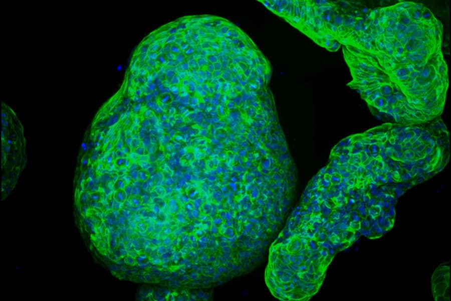
None of this was visible to researchers before because conventional tools destroy the spatial relationships between cells in the process of studying them. Spatial transcriptomics preserves those relationships. The team used a particularly powerful variant called MERFISH, which tracked the activity of 183 different genes across more than 300,000 individual cells, all mapped to their precise locations. The resulting data allowed the researchers to divide the pancreatic tissue into more than 100 distinct spatial domains and analyse which cells inhabited each one and who their neighbours were.
Critically, similar patterns turned up in human pancreatic tissue. Three samples from patients with premalignant lesions but no invasive cancer yet showed comparable cellular niches, with epithelial cells associated with lesions interacting preferentially with immunosuppressive immune cells. The cell types weren’t identical to those in the mouse model — human tissue had a couple of variants not seen in mice — but the fundamental logic appeared conserved. The lesions form niches. The niches corrupt local immunity.
Sharona Tornovsky-Babeay, who was involved in both the data collection and clinical translation of the findings, sees a clear path forward. “Understanding the process of lesion formation and development,” she said, “we may be able to better identify high-risk lesions and, in the future, design strategies that intervene before cancer fully develops.”
That’s the hope, anyway. For now, the findings shift the question of when pancreatic cancer begins to cheat the immune system. The answer appears to be: much earlier than anyone suspected, before the disease is invasive, before it shows on any scan, possibly while the patient is going about their life completely unaware. The immune system isn’t being overwhelmed later. It’s being quietly managed from the very start.
Study link: https://www.gastrojournal.org/article/S0016-5085(25)06655-7/fulltext
ScienceBlog.com has no paywalls, no sponsored content, and no agenda beyond getting the science right. Every story here is written to inform, not to impress an advertiser or push a point of view.
Good science journalism takes time — reading the papers, checking the claims, finding researchers who can put findings in context. We do that work because we think it matters.
If you find this site useful, consider supporting it with a donation. Even a few dollars a month helps keep the coverage independent and free for everyone.